Fergus Tollervey, Manolo U. Rios, Evgenia Zagoriy, Jeffrey B. Woodruff, Julia Mahamid
{"title":"用冷冻电子断层扫描观察秀丽隐杆线虫胚胎中心体的分子结构","authors":"Fergus Tollervey, Manolo U. Rios, Evgenia Zagoriy, Jeffrey B. Woodruff, Julia Mahamid","doi":"10.1016/j.devcel.2024.12.002","DOIUrl":null,"url":null,"abstract":"Centrosomes organize microtubules that are essential for mitotic divisions in animal cells. They consist of centrioles surrounded by pericentriolar material (PCM). Questions related to mechanisms of centriole assembly, PCM organization, and spindle microtubule formation remain unanswered, partly due to limited availability of molecular-resolution structural data inside cells. Here, we use cryo-electron tomography to visualize centrosomes across the cell cycle in cells isolated from <em>C. elegans</em> embryos. We describe a pseudo-timeline of centriole assembly and identify distinct structural features in both mother and daughter centrioles. We find that centrioles and PCM microtubules differ in protofilament number (13 versus 11), which could be explained by atypical γ-tubulin ring complexes with 11-fold symmetry identified at the minus ends of short PCM microtubule segments. We further characterize a porous and disordered network that forms the interconnected PCM. Thus, our work builds a three-dimensional structural atlas that helps explain how centrosomes assemble, grow, and achieve function.","PeriodicalId":11157,"journal":{"name":"Developmental cell","volume":"24 1","pages":""},"PeriodicalIF":8.7000,"publicationDate":"2024-12-24","publicationTypes":"Journal Article","fieldsOfStudy":null,"isOpenAccess":false,"openAccessPdf":"","citationCount":"0","resultStr":"{\"title\":\"Molecular architectures of centrosomes in C. elegans embryos visualized by cryo-electron tomography\",\"authors\":\"Fergus Tollervey, Manolo U. Rios, Evgenia Zagoriy, Jeffrey B. Woodruff, Julia Mahamid\",\"doi\":\"10.1016/j.devcel.2024.12.002\",\"DOIUrl\":null,\"url\":null,\"abstract\":\"Centrosomes organize microtubules that are essential for mitotic divisions in animal cells. They consist of centrioles surrounded by pericentriolar material (PCM). Questions related to mechanisms of centriole assembly, PCM organization, and spindle microtubule formation remain unanswered, partly due to limited availability of molecular-resolution structural data inside cells. Here, we use cryo-electron tomography to visualize centrosomes across the cell cycle in cells isolated from <em>C. elegans</em> embryos. We describe a pseudo-timeline of centriole assembly and identify distinct structural features in both mother and daughter centrioles. We find that centrioles and PCM microtubules differ in protofilament number (13 versus 11), which could be explained by atypical γ-tubulin ring complexes with 11-fold symmetry identified at the minus ends of short PCM microtubule segments. We further characterize a porous and disordered network that forms the interconnected PCM. Thus, our work builds a three-dimensional structural atlas that helps explain how centrosomes assemble, grow, and achieve function.\",\"PeriodicalId\":11157,\"journal\":{\"name\":\"Developmental cell\",\"volume\":\"24 1\",\"pages\":\"\"},\"PeriodicalIF\":8.7000,\"publicationDate\":\"2024-12-24\",\"publicationTypes\":\"Journal Article\",\"fieldsOfStudy\":null,\"isOpenAccess\":false,\"openAccessPdf\":\"\",\"citationCount\":\"0\",\"resultStr\":null,\"platform\":\"Semanticscholar\",\"paperid\":null,\"PeriodicalName\":\"Developmental cell\",\"FirstCategoryId\":\"99\",\"ListUrlMain\":\"https://doi.org/10.1016/j.devcel.2024.12.002\",\"RegionNum\":1,\"RegionCategory\":\"生物学\",\"ArticlePicture\":[],\"TitleCN\":null,\"AbstractTextCN\":null,\"PMCID\":null,\"EPubDate\":\"\",\"PubModel\":\"\",\"JCR\":\"Q1\",\"JCRName\":\"CELL BIOLOGY\",\"Score\":null,\"Total\":0}","platform":"Semanticscholar","paperid":null,"PeriodicalName":"Developmental cell","FirstCategoryId":"99","ListUrlMain":"https://doi.org/10.1016/j.devcel.2024.12.002","RegionNum":1,"RegionCategory":"生物学","ArticlePicture":[],"TitleCN":null,"AbstractTextCN":null,"PMCID":null,"EPubDate":"","PubModel":"","JCR":"Q1","JCRName":"CELL BIOLOGY","Score":null,"Total":0}
Molecular architectures of centrosomes in C. elegans embryos visualized by cryo-electron tomography
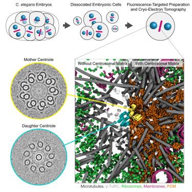
Centrosomes organize microtubules that are essential for mitotic divisions in animal cells. They consist of centrioles surrounded by pericentriolar material (PCM). Questions related to mechanisms of centriole assembly, PCM organization, and spindle microtubule formation remain unanswered, partly due to limited availability of molecular-resolution structural data inside cells. Here, we use cryo-electron tomography to visualize centrosomes across the cell cycle in cells isolated from C. elegans embryos. We describe a pseudo-timeline of centriole assembly and identify distinct structural features in both mother and daughter centrioles. We find that centrioles and PCM microtubules differ in protofilament number (13 versus 11), which could be explained by atypical γ-tubulin ring complexes with 11-fold symmetry identified at the minus ends of short PCM microtubule segments. We further characterize a porous and disordered network that forms the interconnected PCM. Thus, our work builds a three-dimensional structural atlas that helps explain how centrosomes assemble, grow, and achieve function.
期刊介绍:
Developmental Cell, established in 2001, is a comprehensive journal that explores a wide range of topics in cell and developmental biology. Our publication encompasses work across various disciplines within biology, with a particular emphasis on investigating the intersections between cell biology, developmental biology, and other related fields. Our primary objective is to present research conducted through a cell biological perspective, addressing the essential mechanisms governing cell function, cellular interactions, and responses to the environment. Moreover, we focus on understanding the collective behavior of cells, culminating in the formation of tissues, organs, and whole organisms, while also investigating the consequences of any malfunctions in these intricate processes.


 分享
分享
 求助内容:
求助内容: 应助结果提醒方式:
 扫码关注我们
