Luca Giuliani, Carlo Ottonello, Alessandra Giuliani, Lucia Bondì, Paolo Ronconi, Valerio Tempesta, Patrizia Pacini, Vito Cantisani
{"title":"MRI对足底板疾病的评价:“压力测试”的诊断价值。","authors":"Luca Giuliani, Carlo Ottonello, Alessandra Giuliani, Lucia Bondì, Paolo Ronconi, Valerio Tempesta, Patrizia Pacini, Vito Cantisani","doi":"10.1186/s10195-024-00814-x","DOIUrl":null,"url":null,"abstract":"<p><strong>Introduction: </strong>The plantar plate, also called the plantar ligament, is a fibrocartilaginous structure found in the metatarsophalangeal (MTP) and interphalangeal (IP) joints. Our study aimed to evaluate the role of magnetic resonance imaging (MRI) performed with the patient in the standard position or with joint hyperextension (the \"stress test\", ST) in the study of plantar plate (PP) disease that involves metatarsophalangeal joints.</p><p><strong>Materials and methods: </strong>All patients underwent forefoot MRI (Atroscan C, Esaote, Genoa, Italy), operating at 0.2 T. All patients first underwent a standard MRI examination (coronal T1 and T2 weighted image (WI) with fat suppression and axial and sagittal T2 WI); the examination was completed by performing a stress test (hyperextension of toes). The ST is an easy task to perform and is not time-consuming (requiring only one additional sagittal fast spin echo (FSE) T2-weighted MRI sequence; repetition time/ echo time (TR/TE): 3200/90 ms) for patients and operators. A 45°-dorsiflexion ST was performed for approximately 2.30 min, the time required to complete the sequence. No further diagnostic investigations were necessary; no patients underwent arthrography or arthro-MRI. The examinations were performed in a double-blind mode by two operators with proven experience in musculoskeletal radiology; no cases of intra-operator discordance were found.</p><p><strong>Results: </strong>Twenty-five patients were recruited into our study over a 2-year period; 15 were positive for metatarsal pain and 10 were controls. Before treatment (surgery), all patients displaying symptoms underwent evaluation. As a result, the imaging features accurately represented the natural and actual conditions of the lesions. Among the symptomatic patients, 11 out of the 15 exhibited a PP tear or dysfunction in both the standard position and the ST. Additionally, two out of the 15 individuals displayed a tear in the ST alone, with no indication of it in the standard position. In contrast, two out of 15 patients showed no evidence of a PP tear in either the standard position or the ST. However, these two patients demonstrated dorsal subluxation during the ST, likely due to micro-instability resulting from PP failure. In the asymptomatic patients, nine out of the 10 individuals were found to be negative for PP dysfunction. Only one out of the 10 patients exhibited dorsal subluxation solely in the ST, indicative of plantar plate dysfunction, but no evidence of a tear in the PP. In the asymptomatic patients, standard MRI provided a specificity of 100% and a high negative predictive value (NPV) (90%), while the latter increased with the ST (specificity and NPV equal to 100%). In symptomatic patients, standard MRI gave a sensitivity of 75% when assessing a PP tear, which increased to 100% with the ST; the sensitivity of standard MRI the evaluation of MF subluxation was 60%, but it reached 100% with the ST.</p><p><strong>Conclusions: </strong>In our study, by introducing the ST, the sensitivity in both the diagnosis of a PP tear and the evaluation of MTP subluxation reached 100% (a surgical assessment was performed on all positive patients for confirmation). Ultrasound has the advantage of being a non-invasive method. However, comparing the results of our study with the data available in the literature, ultrasound has a lower sensitivity and a negative predictive value. Also, ultrasound does not allow for the assessment of possible bone marrow oedema or the degree of concomitant arthritis. If other studies in the literature confirm these results, it will be possible to consider incorporating the ST into diagnostic practice in the future.</p>","PeriodicalId":48603,"journal":{"name":"Journal of Orthopaedics and Traumatology","volume":"25 1","pages":"70"},"PeriodicalIF":3.7000,"publicationDate":"2024-12-24","publicationTypes":"Journal Article","fieldsOfStudy":null,"isOpenAccess":false,"openAccessPdf":"https://www.ncbi.nlm.nih.gov/pmc/articles/PMC11668703/pdf/","citationCount":"0","resultStr":"{\"title\":\"MRI in the evaluation of plantar plate disease: diagnostic value of the \\\"stress test\\\".\",\"authors\":\"Luca Giuliani, Carlo Ottonello, Alessandra Giuliani, Lucia Bondì, Paolo Ronconi, Valerio Tempesta, Patrizia Pacini, Vito Cantisani\",\"doi\":\"10.1186/s10195-024-00814-x\",\"DOIUrl\":null,\"url\":null,\"abstract\":\"<p><strong>Introduction: </strong>The plantar plate, also called the plantar ligament, is a fibrocartilaginous structure found in the metatarsophalangeal (MTP) and interphalangeal (IP) joints. Our study aimed to evaluate the role of magnetic resonance imaging (MRI) performed with the patient in the standard position or with joint hyperextension (the \\\"stress test\\\", ST) in the study of plantar plate (PP) disease that involves metatarsophalangeal joints.</p><p><strong>Materials and methods: </strong>All patients underwent forefoot MRI (Atroscan C, Esaote, Genoa, Italy), operating at 0.2 T. All patients first underwent a standard MRI examination (coronal T1 and T2 weighted image (WI) with fat suppression and axial and sagittal T2 WI); the examination was completed by performing a stress test (hyperextension of toes). The ST is an easy task to perform and is not time-consuming (requiring only one additional sagittal fast spin echo (FSE) T2-weighted MRI sequence; repetition time/ echo time (TR/TE): 3200/90 ms) for patients and operators. A 45°-dorsiflexion ST was performed for approximately 2.30 min, the time required to complete the sequence. No further diagnostic investigations were necessary; no patients underwent arthrography or arthro-MRI. The examinations were performed in a double-blind mode by two operators with proven experience in musculoskeletal radiology; no cases of intra-operator discordance were found.</p><p><strong>Results: </strong>Twenty-five patients were recruited into our study over a 2-year period; 15 were positive for metatarsal pain and 10 were controls. Before treatment (surgery), all patients displaying symptoms underwent evaluation. As a result, the imaging features accurately represented the natural and actual conditions of the lesions. Among the symptomatic patients, 11 out of the 15 exhibited a PP tear or dysfunction in both the standard position and the ST. Additionally, two out of the 15 individuals displayed a tear in the ST alone, with no indication of it in the standard position. In contrast, two out of 15 patients showed no evidence of a PP tear in either the standard position or the ST. However, these two patients demonstrated dorsal subluxation during the ST, likely due to micro-instability resulting from PP failure. In the asymptomatic patients, nine out of the 10 individuals were found to be negative for PP dysfunction. Only one out of the 10 patients exhibited dorsal subluxation solely in the ST, indicative of plantar plate dysfunction, but no evidence of a tear in the PP. In the asymptomatic patients, standard MRI provided a specificity of 100% and a high negative predictive value (NPV) (90%), while the latter increased with the ST (specificity and NPV equal to 100%). In symptomatic patients, standard MRI gave a sensitivity of 75% when assessing a PP tear, which increased to 100% with the ST; the sensitivity of standard MRI the evaluation of MF subluxation was 60%, but it reached 100% with the ST.</p><p><strong>Conclusions: </strong>In our study, by introducing the ST, the sensitivity in both the diagnosis of a PP tear and the evaluation of MTP subluxation reached 100% (a surgical assessment was performed on all positive patients for confirmation). Ultrasound has the advantage of being a non-invasive method. However, comparing the results of our study with the data available in the literature, ultrasound has a lower sensitivity and a negative predictive value. Also, ultrasound does not allow for the assessment of possible bone marrow oedema or the degree of concomitant arthritis. If other studies in the literature confirm these results, it will be possible to consider incorporating the ST into diagnostic practice in the future.</p>\",\"PeriodicalId\":48603,\"journal\":{\"name\":\"Journal of Orthopaedics and Traumatology\",\"volume\":\"25 1\",\"pages\":\"70\"},\"PeriodicalIF\":3.7000,\"publicationDate\":\"2024-12-24\",\"publicationTypes\":\"Journal Article\",\"fieldsOfStudy\":null,\"isOpenAccess\":false,\"openAccessPdf\":\"https://www.ncbi.nlm.nih.gov/pmc/articles/PMC11668703/pdf/\",\"citationCount\":\"0\",\"resultStr\":null,\"platform\":\"Semanticscholar\",\"paperid\":null,\"PeriodicalName\":\"Journal of Orthopaedics and Traumatology\",\"FirstCategoryId\":\"3\",\"ListUrlMain\":\"https://doi.org/10.1186/s10195-024-00814-x\",\"RegionNum\":2,\"RegionCategory\":\"医学\",\"ArticlePicture\":[],\"TitleCN\":null,\"AbstractTextCN\":null,\"PMCID\":null,\"EPubDate\":\"\",\"PubModel\":\"\",\"JCR\":\"Q1\",\"JCRName\":\"ORTHOPEDICS\",\"Score\":null,\"Total\":0}","platform":"Semanticscholar","paperid":null,"PeriodicalName":"Journal of Orthopaedics and Traumatology","FirstCategoryId":"3","ListUrlMain":"https://doi.org/10.1186/s10195-024-00814-x","RegionNum":2,"RegionCategory":"医学","ArticlePicture":[],"TitleCN":null,"AbstractTextCN":null,"PMCID":null,"EPubDate":"","PubModel":"","JCR":"Q1","JCRName":"ORTHOPEDICS","Score":null,"Total":0}
MRI in the evaluation of plantar plate disease: diagnostic value of the "stress test".
Introduction: The plantar plate, also called the plantar ligament, is a fibrocartilaginous structure found in the metatarsophalangeal (MTP) and interphalangeal (IP) joints. Our study aimed to evaluate the role of magnetic resonance imaging (MRI) performed with the patient in the standard position or with joint hyperextension (the "stress test", ST) in the study of plantar plate (PP) disease that involves metatarsophalangeal joints.
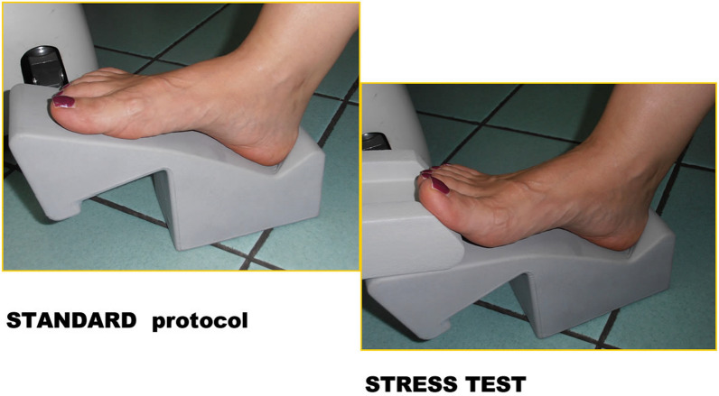
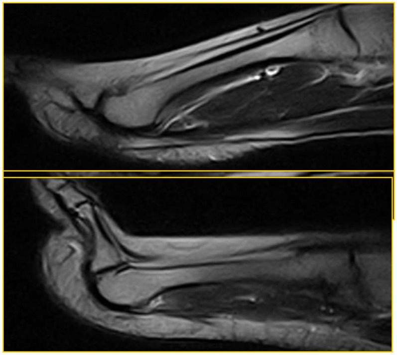
Materials and methods: All patients underwent forefoot MRI (Atroscan C, Esaote, Genoa, Italy), operating at 0.2 T. All patients first underwent a standard MRI examination (coronal T1 and T2 weighted image (WI) with fat suppression and axial and sagittal T2 WI); the examination was completed by performing a stress test (hyperextension of toes). The ST is an easy task to perform and is not time-consuming (requiring only one additional sagittal fast spin echo (FSE) T2-weighted MRI sequence; repetition time/ echo time (TR/TE): 3200/90 ms) for patients and operators. A 45°-dorsiflexion ST was performed for approximately 2.30 min, the time required to complete the sequence. No further diagnostic investigations were necessary; no patients underwent arthrography or arthro-MRI. The examinations were performed in a double-blind mode by two operators with proven experience in musculoskeletal radiology; no cases of intra-operator discordance were found.
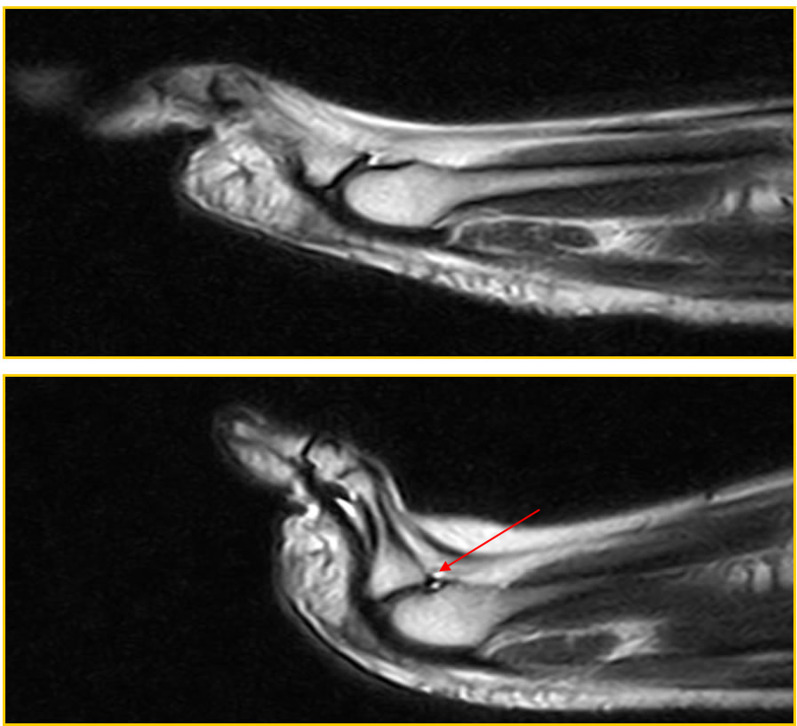
Results: Twenty-five patients were recruited into our study over a 2-year period; 15 were positive for metatarsal pain and 10 were controls. Before treatment (surgery), all patients displaying symptoms underwent evaluation. As a result, the imaging features accurately represented the natural and actual conditions of the lesions. Among the symptomatic patients, 11 out of the 15 exhibited a PP tear or dysfunction in both the standard position and the ST. Additionally, two out of the 15 individuals displayed a tear in the ST alone, with no indication of it in the standard position. In contrast, two out of 15 patients showed no evidence of a PP tear in either the standard position or the ST. However, these two patients demonstrated dorsal subluxation during the ST, likely due to micro-instability resulting from PP failure. In the asymptomatic patients, nine out of the 10 individuals were found to be negative for PP dysfunction. Only one out of the 10 patients exhibited dorsal subluxation solely in the ST, indicative of plantar plate dysfunction, but no evidence of a tear in the PP. In the asymptomatic patients, standard MRI provided a specificity of 100% and a high negative predictive value (NPV) (90%), while the latter increased with the ST (specificity and NPV equal to 100%). In symptomatic patients, standard MRI gave a sensitivity of 75% when assessing a PP tear, which increased to 100% with the ST; the sensitivity of standard MRI the evaluation of MF subluxation was 60%, but it reached 100% with the ST.
Conclusions: In our study, by introducing the ST, the sensitivity in both the diagnosis of a PP tear and the evaluation of MTP subluxation reached 100% (a surgical assessment was performed on all positive patients for confirmation). Ultrasound has the advantage of being a non-invasive method. However, comparing the results of our study with the data available in the literature, ultrasound has a lower sensitivity and a negative predictive value. Also, ultrasound does not allow for the assessment of possible bone marrow oedema or the degree of concomitant arthritis. If other studies in the literature confirm these results, it will be possible to consider incorporating the ST into diagnostic practice in the future.
期刊介绍:
The Journal of Orthopaedics and Traumatology, the official open access peer-reviewed journal of the Italian Society of Orthopaedics and Traumatology, publishes original papers reporting basic or clinical research in the field of orthopaedic and traumatologic surgery, as well as systematic reviews, brief communications, case reports and letters to the Editor. Narrative instructional reviews and commentaries to original articles may be commissioned by Editors from eminent colleagues. The Journal of Orthopaedics and Traumatology aims to be an international forum for the communication and exchange of ideas concerning the various aspects of orthopaedics and musculoskeletal trauma.


 分享
分享
 求助内容:
求助内容: 应助结果提醒方式:
 扫码关注我们
