Dennisha P King, Miral Abdalaziz, Ania K Majewska, Judy L Cameron, Julie L Fudge
{"title":"灵长类动物杏仁核发育中的小胶质细胞形态及早期生活应激的影响。","authors":"Dennisha P King, Miral Abdalaziz, Ania K Majewska, Judy L Cameron, Julie L Fudge","doi":"10.1523/ENEURO.0466-24.2024","DOIUrl":null,"url":null,"abstract":"<p><p>A unique pool of immature glutamatergic neurons in the primate amygdala, known as the paralaminar nucleus (PL), are maturing between infancy and adolescence. The PL is a potential substrate for the steep growth curve of amygdala volume during this developmental period. A microglial component is also embedded among the PL neurons and likely supports local neuronal maturation and emerging synaptogenesis. Microglia may alter neuronal growth following environmental perturbations such as stress. Using multiple measures in rhesus macaques, we found that microglia in the infant primate PL had relatively large somas and a small arbor size. In contrast, microglia in the adolescent PL had a smaller soma and a larger dendritic arbor. We then examined microglial morphology in the PL after a novel maternal separation protocol, to examine the effects of early life stress. After maternal separation, the microglia had increased soma size, arbor size, and complexity. Surprisingly, strong effects were seen not only in the infant PL, but also in the adolescent PL from subjects who had experienced the separation many years earlier. We conclude that under normal maternal-rearing conditions, PL microglia morphology tracks PL neuronal growth, progressing to a more \"mature\" phenotype by adolescence. Maternal separation has long-lasting effects on microglia, altering their normal developmental trajectory, and resulting in a \"hyper-ramified\" phenotype that persists for years. We speculate that these changes have consequences for neuronal development in young primates.</p>","PeriodicalId":11617,"journal":{"name":"eNeuro","volume":" ","pages":""},"PeriodicalIF":2.6000,"publicationDate":"2025-01-15","publicationTypes":"Journal Article","fieldsOfStudy":null,"isOpenAccess":false,"openAccessPdf":"https://www.ncbi.nlm.nih.gov/pmc/articles/PMC11735683/pdf/","citationCount":"0","resultStr":"{\"title\":\"Microglia Morphology in the Developing Primate Amygdala and Effects of Early Life Stress.\",\"authors\":\"Dennisha P King, Miral Abdalaziz, Ania K Majewska, Judy L Cameron, Julie L Fudge\",\"doi\":\"10.1523/ENEURO.0466-24.2024\",\"DOIUrl\":null,\"url\":null,\"abstract\":\"<p><p>A unique pool of immature glutamatergic neurons in the primate amygdala, known as the paralaminar nucleus (PL), are maturing between infancy and adolescence. The PL is a potential substrate for the steep growth curve of amygdala volume during this developmental period. A microglial component is also embedded among the PL neurons and likely supports local neuronal maturation and emerging synaptogenesis. Microglia may alter neuronal growth following environmental perturbations such as stress. Using multiple measures in rhesus macaques, we found that microglia in the infant primate PL had relatively large somas and a small arbor size. In contrast, microglia in the adolescent PL had a smaller soma and a larger dendritic arbor. We then examined microglial morphology in the PL after a novel maternal separation protocol, to examine the effects of early life stress. After maternal separation, the microglia had increased soma size, arbor size, and complexity. Surprisingly, strong effects were seen not only in the infant PL, but also in the adolescent PL from subjects who had experienced the separation many years earlier. We conclude that under normal maternal-rearing conditions, PL microglia morphology tracks PL neuronal growth, progressing to a more \\\"mature\\\" phenotype by adolescence. Maternal separation has long-lasting effects on microglia, altering their normal developmental trajectory, and resulting in a \\\"hyper-ramified\\\" phenotype that persists for years. We speculate that these changes have consequences for neuronal development in young primates.</p>\",\"PeriodicalId\":11617,\"journal\":{\"name\":\"eNeuro\",\"volume\":\" \",\"pages\":\"\"},\"PeriodicalIF\":2.6000,\"publicationDate\":\"2025-01-15\",\"publicationTypes\":\"Journal Article\",\"fieldsOfStudy\":null,\"isOpenAccess\":false,\"openAccessPdf\":\"https://www.ncbi.nlm.nih.gov/pmc/articles/PMC11735683/pdf/\",\"citationCount\":\"0\",\"resultStr\":null,\"platform\":\"Semanticscholar\",\"paperid\":null,\"PeriodicalName\":\"eNeuro\",\"FirstCategoryId\":\"3\",\"ListUrlMain\":\"https://doi.org/10.1523/ENEURO.0466-24.2024\",\"RegionNum\":3,\"RegionCategory\":\"医学\",\"ArticlePicture\":[],\"TitleCN\":null,\"AbstractTextCN\":null,\"PMCID\":null,\"EPubDate\":\"2025/1/1 0:00:00\",\"PubModel\":\"Print\",\"JCR\":\"Q3\",\"JCRName\":\"NEUROSCIENCES\",\"Score\":null,\"Total\":0}","platform":"Semanticscholar","paperid":null,"PeriodicalName":"eNeuro","FirstCategoryId":"3","ListUrlMain":"https://doi.org/10.1523/ENEURO.0466-24.2024","RegionNum":3,"RegionCategory":"医学","ArticlePicture":[],"TitleCN":null,"AbstractTextCN":null,"PMCID":null,"EPubDate":"2025/1/1 0:00:00","PubModel":"Print","JCR":"Q3","JCRName":"NEUROSCIENCES","Score":null,"Total":0}
Microglia Morphology in the Developing Primate Amygdala and Effects of Early Life Stress.
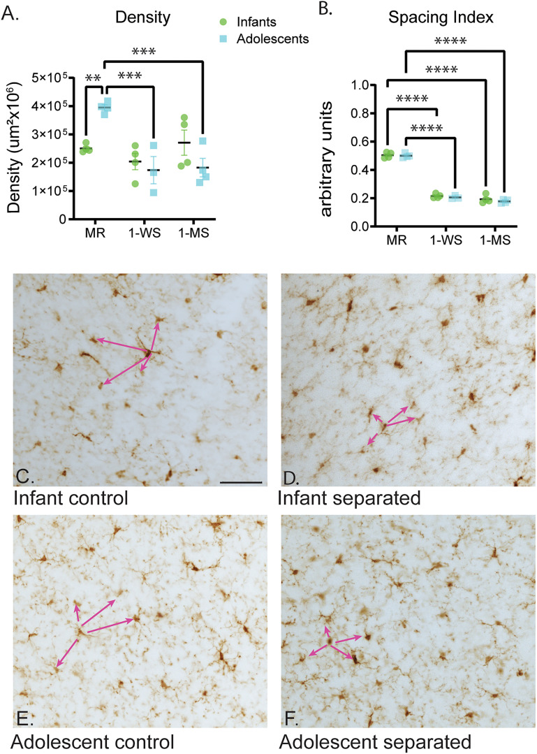
A unique pool of immature glutamatergic neurons in the primate amygdala, known as the paralaminar nucleus (PL), are maturing between infancy and adolescence. The PL is a potential substrate for the steep growth curve of amygdala volume during this developmental period. A microglial component is also embedded among the PL neurons and likely supports local neuronal maturation and emerging synaptogenesis. Microglia may alter neuronal growth following environmental perturbations such as stress. Using multiple measures in rhesus macaques, we found that microglia in the infant primate PL had relatively large somas and a small arbor size. In contrast, microglia in the adolescent PL had a smaller soma and a larger dendritic arbor. We then examined microglial morphology in the PL after a novel maternal separation protocol, to examine the effects of early life stress. After maternal separation, the microglia had increased soma size, arbor size, and complexity. Surprisingly, strong effects were seen not only in the infant PL, but also in the adolescent PL from subjects who had experienced the separation many years earlier. We conclude that under normal maternal-rearing conditions, PL microglia morphology tracks PL neuronal growth, progressing to a more "mature" phenotype by adolescence. Maternal separation has long-lasting effects on microglia, altering their normal developmental trajectory, and resulting in a "hyper-ramified" phenotype that persists for years. We speculate that these changes have consequences for neuronal development in young primates.
期刊介绍:
An open-access journal from the Society for Neuroscience, eNeuro publishes high-quality, broad-based, peer-reviewed research focused solely on the field of neuroscience. eNeuro embodies an emerging scientific vision that offers a new experience for authors and readers, all in support of the Society’s mission to advance understanding of the brain and nervous system.


 分享
分享
 求助内容:
求助内容: 应助结果提醒方式:
 扫码关注我们
