Sarah R. Stevenson, Svetomir B. Tzokov, Indrajit Lahiri, Kathryn R. Ayscough, Per A. Bullough
{"title":"酵母adp -肌动蛋白丝在2.5 Å分辨率下的低温电镜重建。与脊椎动物f -肌动蛋白的比较","authors":"Sarah R. Stevenson, Svetomir B. Tzokov, Indrajit Lahiri, Kathryn R. Ayscough, Per A. Bullough","doi":"10.1016/j.str.2024.12.008","DOIUrl":null,"url":null,"abstract":"The core component of the actin cytoskeleton is the globular protein G-actin, which reversibly polymerizes into filaments (F-actin). Budding yeast possesses a single actin that shares 87%–89% sequence identity with vertebrate actin isoforms. Previous structural studies indicate very close overlap of main-chain backbones. Intriguingly, however, substitution of yeast <em>ACT1</em> with vertebrate β-cytoplasmic actin severely disrupts cell function and the substitution with a skeletal muscle isoform is lethal. Here we report a 2.5 Å structure of budding yeast F-actin. Previously unresolved side-chain information allows us to highlight four main differences in the comparison of yeast and vertebrate ADP F-actins: a more open nucleotide binding pocket; a more solvent exposed C-terminus; a rearrangement of inter-subunit binding interactions in the vicinity of the D loop and changes in the hydrogen bonding network in the vicinity of histidine 73 (yeast actin) and methyl-histidine 73 (vertebrate actin).","PeriodicalId":22168,"journal":{"name":"Structure","volume":"8 1","pages":""},"PeriodicalIF":4.3000,"publicationDate":"2025-01-10","publicationTypes":"Journal Article","fieldsOfStudy":null,"isOpenAccess":false,"openAccessPdf":"","citationCount":"0","resultStr":"{\"title\":\"Cryo-EM reconstruction of yeast ADP-actin filament at 2.5 Å resolution. A comparison with vertebrate F-actin\",\"authors\":\"Sarah R. Stevenson, Svetomir B. Tzokov, Indrajit Lahiri, Kathryn R. Ayscough, Per A. Bullough\",\"doi\":\"10.1016/j.str.2024.12.008\",\"DOIUrl\":null,\"url\":null,\"abstract\":\"The core component of the actin cytoskeleton is the globular protein G-actin, which reversibly polymerizes into filaments (F-actin). Budding yeast possesses a single actin that shares 87%–89% sequence identity with vertebrate actin isoforms. Previous structural studies indicate very close overlap of main-chain backbones. Intriguingly, however, substitution of yeast <em>ACT1</em> with vertebrate β-cytoplasmic actin severely disrupts cell function and the substitution with a skeletal muscle isoform is lethal. Here we report a 2.5 Å structure of budding yeast F-actin. Previously unresolved side-chain information allows us to highlight four main differences in the comparison of yeast and vertebrate ADP F-actins: a more open nucleotide binding pocket; a more solvent exposed C-terminus; a rearrangement of inter-subunit binding interactions in the vicinity of the D loop and changes in the hydrogen bonding network in the vicinity of histidine 73 (yeast actin) and methyl-histidine 73 (vertebrate actin).\",\"PeriodicalId\":22168,\"journal\":{\"name\":\"Structure\",\"volume\":\"8 1\",\"pages\":\"\"},\"PeriodicalIF\":4.3000,\"publicationDate\":\"2025-01-10\",\"publicationTypes\":\"Journal Article\",\"fieldsOfStudy\":null,\"isOpenAccess\":false,\"openAccessPdf\":\"\",\"citationCount\":\"0\",\"resultStr\":null,\"platform\":\"Semanticscholar\",\"paperid\":null,\"PeriodicalName\":\"Structure\",\"FirstCategoryId\":\"99\",\"ListUrlMain\":\"https://doi.org/10.1016/j.str.2024.12.008\",\"RegionNum\":2,\"RegionCategory\":\"生物学\",\"ArticlePicture\":[],\"TitleCN\":null,\"AbstractTextCN\":null,\"PMCID\":null,\"EPubDate\":\"\",\"PubModel\":\"\",\"JCR\":\"Q2\",\"JCRName\":\"BIOCHEMISTRY & MOLECULAR BIOLOGY\",\"Score\":null,\"Total\":0}","platform":"Semanticscholar","paperid":null,"PeriodicalName":"Structure","FirstCategoryId":"99","ListUrlMain":"https://doi.org/10.1016/j.str.2024.12.008","RegionNum":2,"RegionCategory":"生物学","ArticlePicture":[],"TitleCN":null,"AbstractTextCN":null,"PMCID":null,"EPubDate":"","PubModel":"","JCR":"Q2","JCRName":"BIOCHEMISTRY & MOLECULAR BIOLOGY","Score":null,"Total":0}
引用次数: 0
摘要
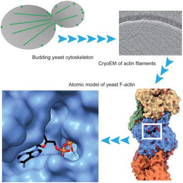
肌动蛋白细胞骨架的核心成分是球形蛋白g -肌动蛋白,它可逆地聚合成细丝(f -肌动蛋白)。出芽酵母具有一个单一的肌动蛋白,与脊椎动物肌动蛋白同工型具有87%-89%的序列一致性。先前的结构研究表明,主链主干的重叠非常紧密。然而,有趣的是,用脊椎动物β-细胞质肌动蛋白替代酵母ACT1严重破坏细胞功能,并用骨骼肌同种异构体替代是致命的。在这里,我们报告了出芽酵母f -肌动蛋白的2.5 Å结构。先前未解决的侧链信息使我们能够突出酵母和脊椎动物ADP f -actin比较中的四个主要差异:更开放的核苷酸结合袋;一个更具溶剂性的暴露c端;D环附近亚基间结合相互作用的重排以及组氨酸73(酵母肌动蛋白)和甲基组氨酸73(脊椎动物肌动蛋白)附近氢键网络的变化。
Cryo-EM reconstruction of yeast ADP-actin filament at 2.5 Å resolution. A comparison with vertebrate F-actin
The core component of the actin cytoskeleton is the globular protein G-actin, which reversibly polymerizes into filaments (F-actin). Budding yeast possesses a single actin that shares 87%–89% sequence identity with vertebrate actin isoforms. Previous structural studies indicate very close overlap of main-chain backbones. Intriguingly, however, substitution of yeast ACT1 with vertebrate β-cytoplasmic actin severely disrupts cell function and the substitution with a skeletal muscle isoform is lethal. Here we report a 2.5 Å structure of budding yeast F-actin. Previously unresolved side-chain information allows us to highlight four main differences in the comparison of yeast and vertebrate ADP F-actins: a more open nucleotide binding pocket; a more solvent exposed C-terminus; a rearrangement of inter-subunit binding interactions in the vicinity of the D loop and changes in the hydrogen bonding network in the vicinity of histidine 73 (yeast actin) and methyl-histidine 73 (vertebrate actin).
期刊介绍:
Structure aims to publish papers of exceptional interest in the field of structural biology. The journal strives to be essential reading for structural biologists, as well as biologists and biochemists that are interested in macromolecular structure and function. Structure strongly encourages the submission of manuscripts that present structural and molecular insights into biological function and mechanism. Other reports that address fundamental questions in structural biology, such as structure-based examinations of protein evolution, folding, and/or design, will also be considered. We will consider the application of any method, experimental or computational, at high or low resolution, to conduct structural investigations, as long as the method is appropriate for the biological, functional, and mechanistic question(s) being addressed. Likewise, reports describing single-molecule analysis of biological mechanisms are welcome.
In general, the editors encourage submission of experimental structural studies that are enriched by an analysis of structure-activity relationships and will not consider studies that solely report structural information unless the structure or analysis is of exceptional and broad interest. Studies reporting only homology models, de novo models, or molecular dynamics simulations are also discouraged unless the models are informed by or validated by novel experimental data; rationalization of a large body of existing experimental evidence and making testable predictions based on a model or simulation is often not considered sufficient.


 分享
分享
 求助内容:
求助内容: 应助结果提醒方式:
 扫码关注我们
