Yuehan Ni, Jiamiao Wu, Fengqi Liu, Yating Yi, Xiangjiao Meng, Xiang Gao, Luyi Xiao, Weiwei Zhou, Zexi Chen, Peng Chu, Dan Xing, Ye Yuan, Donghui Ding, Ge Shen, Min Yang, Ronjie Wu, Ling Wang, Luiza Martins Nascentes Melo, Sien Lin, Xiaoguang Cheng, Gang Li, Alpaslan Tasdogan, Jessalyn M. Ubellacker, Hu Zhao, Shentong Fang, Bo Shen
{"title":"LepR+基质细胞在光学清除的小鼠骨半切面的深度成像","authors":"Yuehan Ni, Jiamiao Wu, Fengqi Liu, Yating Yi, Xiangjiao Meng, Xiang Gao, Luyi Xiao, Weiwei Zhou, Zexi Chen, Peng Chu, Dan Xing, Ye Yuan, Donghui Ding, Ge Shen, Min Yang, Ronjie Wu, Ling Wang, Luiza Martins Nascentes Melo, Sien Lin, Xiaoguang Cheng, Gang Li, Alpaslan Tasdogan, Jessalyn M. Ubellacker, Hu Zhao, Shentong Fang, Bo Shen","doi":"10.1038/s41413-024-00387-9","DOIUrl":null,"url":null,"abstract":"<p>Tissue clearing combined with high-resolution confocal imaging is a cutting-edge approach for dissecting the three-dimensional (3D) architecture of tissues and deciphering cellular spatial interactions under physiological and pathological conditions. Deciphering the spatial interaction of leptin receptor-expressing (LepR<sup>+</sup>) stromal cells with other compartments in the bone marrow is crucial for a deeper understanding of the stem cell niche and the skeletal tissue. In this study, we introduce an optimized protocol for the 3D analysis of skeletal tissues, enabling the visualization of hematopoietic and stromal cells, especially LepR<sup>+</sup> stromal cells, within optically cleared bone hemisections. Our method preserves the 3D tissue architecture and is extendable to other hematopoietic sites such as calvaria and vertebrae. The protocol entails tissue fixation, decalcification, and cryosectioning to reveal the marrow cavity. Completed within approximately 12 days, this process yields highly transparent tissues that maintain genetically encoded or antibody-stained fluorescent signals. The bone hemisections are compatible with diverse antibody labeling strategies. Confocal microscopy of these transparent samples allows for qualitative and quantitative image analysis using Aivia or Bitplane Imaris software, assessing a spectrum of parameters. With proper storage, the fluorescent signal in the stained and cleared bone hemisections remains intact for at least 2–3 months. This protocol is robust, straightforward to implement, and highly reproducible, offering a valuable tool for tissue architecture and cellular interaction studies.</p>","PeriodicalId":9134,"journal":{"name":"Bone Research","volume":"15 1","pages":""},"PeriodicalIF":15.0000,"publicationDate":"2025-01-13","publicationTypes":"Journal Article","fieldsOfStudy":null,"isOpenAccess":false,"openAccessPdf":"","citationCount":"0","resultStr":"{\"title\":\"Deep imaging of LepR+ stromal cells in optically cleared murine bone hemisections\",\"authors\":\"Yuehan Ni, Jiamiao Wu, Fengqi Liu, Yating Yi, Xiangjiao Meng, Xiang Gao, Luyi Xiao, Weiwei Zhou, Zexi Chen, Peng Chu, Dan Xing, Ye Yuan, Donghui Ding, Ge Shen, Min Yang, Ronjie Wu, Ling Wang, Luiza Martins Nascentes Melo, Sien Lin, Xiaoguang Cheng, Gang Li, Alpaslan Tasdogan, Jessalyn M. Ubellacker, Hu Zhao, Shentong Fang, Bo Shen\",\"doi\":\"10.1038/s41413-024-00387-9\",\"DOIUrl\":null,\"url\":null,\"abstract\":\"<p>Tissue clearing combined with high-resolution confocal imaging is a cutting-edge approach for dissecting the three-dimensional (3D) architecture of tissues and deciphering cellular spatial interactions under physiological and pathological conditions. Deciphering the spatial interaction of leptin receptor-expressing (LepR<sup>+</sup>) stromal cells with other compartments in the bone marrow is crucial for a deeper understanding of the stem cell niche and the skeletal tissue. In this study, we introduce an optimized protocol for the 3D analysis of skeletal tissues, enabling the visualization of hematopoietic and stromal cells, especially LepR<sup>+</sup> stromal cells, within optically cleared bone hemisections. Our method preserves the 3D tissue architecture and is extendable to other hematopoietic sites such as calvaria and vertebrae. The protocol entails tissue fixation, decalcification, and cryosectioning to reveal the marrow cavity. Completed within approximately 12 days, this process yields highly transparent tissues that maintain genetically encoded or antibody-stained fluorescent signals. The bone hemisections are compatible with diverse antibody labeling strategies. Confocal microscopy of these transparent samples allows for qualitative and quantitative image analysis using Aivia or Bitplane Imaris software, assessing a spectrum of parameters. With proper storage, the fluorescent signal in the stained and cleared bone hemisections remains intact for at least 2–3 months. This protocol is robust, straightforward to implement, and highly reproducible, offering a valuable tool for tissue architecture and cellular interaction studies.</p>\",\"PeriodicalId\":9134,\"journal\":{\"name\":\"Bone Research\",\"volume\":\"15 1\",\"pages\":\"\"},\"PeriodicalIF\":15.0000,\"publicationDate\":\"2025-01-13\",\"publicationTypes\":\"Journal Article\",\"fieldsOfStudy\":null,\"isOpenAccess\":false,\"openAccessPdf\":\"\",\"citationCount\":\"0\",\"resultStr\":null,\"platform\":\"Semanticscholar\",\"paperid\":null,\"PeriodicalName\":\"Bone Research\",\"FirstCategoryId\":\"3\",\"ListUrlMain\":\"https://doi.org/10.1038/s41413-024-00387-9\",\"RegionNum\":1,\"RegionCategory\":\"医学\",\"ArticlePicture\":[],\"TitleCN\":null,\"AbstractTextCN\":null,\"PMCID\":null,\"EPubDate\":\"\",\"PubModel\":\"\",\"JCR\":\"Q1\",\"JCRName\":\"CELL & TISSUE ENGINEERING\",\"Score\":null,\"Total\":0}","platform":"Semanticscholar","paperid":null,"PeriodicalName":"Bone Research","FirstCategoryId":"3","ListUrlMain":"https://doi.org/10.1038/s41413-024-00387-9","RegionNum":1,"RegionCategory":"医学","ArticlePicture":[],"TitleCN":null,"AbstractTextCN":null,"PMCID":null,"EPubDate":"","PubModel":"","JCR":"Q1","JCRName":"CELL & TISSUE ENGINEERING","Score":null,"Total":0}
Deep imaging of LepR+ stromal cells in optically cleared murine bone hemisections
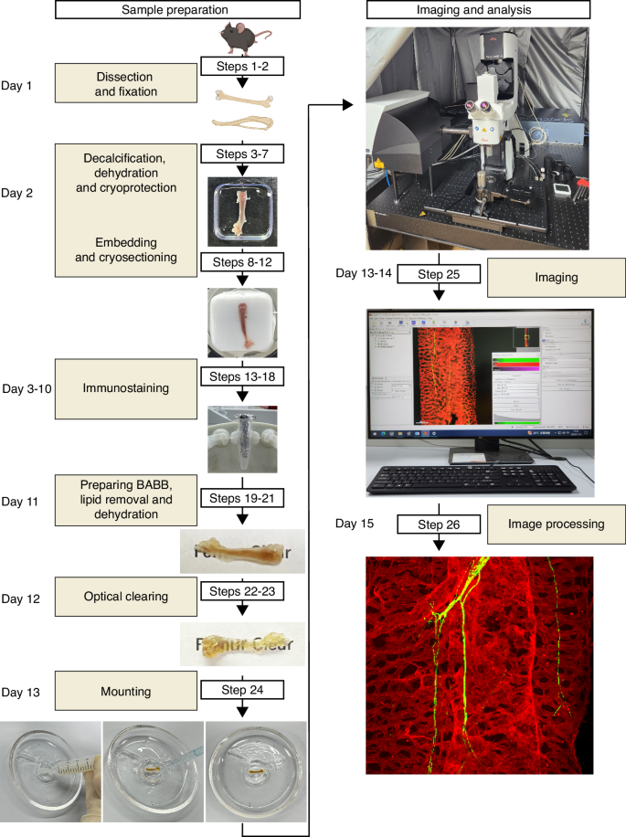
Tissue clearing combined with high-resolution confocal imaging is a cutting-edge approach for dissecting the three-dimensional (3D) architecture of tissues and deciphering cellular spatial interactions under physiological and pathological conditions. Deciphering the spatial interaction of leptin receptor-expressing (LepR+) stromal cells with other compartments in the bone marrow is crucial for a deeper understanding of the stem cell niche and the skeletal tissue. In this study, we introduce an optimized protocol for the 3D analysis of skeletal tissues, enabling the visualization of hematopoietic and stromal cells, especially LepR+ stromal cells, within optically cleared bone hemisections. Our method preserves the 3D tissue architecture and is extendable to other hematopoietic sites such as calvaria and vertebrae. The protocol entails tissue fixation, decalcification, and cryosectioning to reveal the marrow cavity. Completed within approximately 12 days, this process yields highly transparent tissues that maintain genetically encoded or antibody-stained fluorescent signals. The bone hemisections are compatible with diverse antibody labeling strategies. Confocal microscopy of these transparent samples allows for qualitative and quantitative image analysis using Aivia or Bitplane Imaris software, assessing a spectrum of parameters. With proper storage, the fluorescent signal in the stained and cleared bone hemisections remains intact for at least 2–3 months. This protocol is robust, straightforward to implement, and highly reproducible, offering a valuable tool for tissue architecture and cellular interaction studies.
期刊介绍:
Established in 2013, Bone Research is a newly-founded English-language periodical that centers on the basic and clinical facets of bone biology, pathophysiology, and regeneration. It is dedicated to championing key findings emerging from both basic investigations and clinical research concerning bone-related topics. The journal's objective is to globally disseminate research in bone-related physiology, pathology, diseases, and treatment, contributing to the advancement of knowledge in this field.


 分享
分享
 求助内容:
求助内容: 应助结果提醒方式:
 扫码关注我们
