Luca Bernecker, Ellisiv B Mathiesen, Tor Ingebrigtsen, Jørgen Isaksen, Liv-Hege Johnsen, Torgil Riise Vangberg
{"title":"基于贴片的深度学习颅内狭窄和动脉瘤检测方法——Tromsø研究。","authors":"Luca Bernecker, Ellisiv B Mathiesen, Tor Ingebrigtsen, Jørgen Isaksen, Liv-Hege Johnsen, Torgil Riise Vangberg","doi":"10.1007/s12021-024-09697-z","DOIUrl":null,"url":null,"abstract":"<p><p>Intracranial atherosclerotic stenosis (ICAS) and intracranial aneurysms are prevalent conditions in the cerebrovascular system. ICAS causes a narrowing of the arterial lumen, thereby restricting blood flow, while aneurysms involve the ballooning of blood vessels. Both conditions can lead to severe outcomes, such as stroke or vessel rupture, which can be fatal. Early detection is crucial for effective intervention. In this study, we introduced a method that combines classical computer vision techniques with deep learning to detect intracranial aneurysms and ICAS in time-of-flight magnetic resonance angiography images. The process began with skull-stripping, followed by an affine transformation to align the images to a common atlas space. We then focused on the region of interest, including the circle of Willis, by cropping the relevant area. A segmentation algorithm was used to isolate the arteries, after which a patch-wise residual neural network was applied across the image. A voting mechanism was then employed to identify the presence of atrophies. Our method achieved accuracies of 76.5% for aneurysms and 82.4% for ICAS. Notably, when occlusions were not considered, the accuracy for ICAS detection improved to 85.7%. While the algorithm performed well for localized pathological findings, it was less effective at detecting occlusions, which involved long-range dependencies in the MRIs. This limitation was due to the architectural design of the patch-wise deep learning approach. Regardless, this can, in the future, be mitigated in a multi-scale patch-wise algorithm.</p>","PeriodicalId":49761,"journal":{"name":"Neuroinformatics","volume":"23 1","pages":"8"},"PeriodicalIF":3.1000,"publicationDate":"2025-01-15","publicationTypes":"Journal Article","fieldsOfStudy":null,"isOpenAccess":false,"openAccessPdf":"https://www.ncbi.nlm.nih.gov/pmc/articles/PMC11735523/pdf/","citationCount":"0","resultStr":"{\"title\":\"Patch-Wise Deep Learning Method for Intracranial Stenosis and Aneurysm Detection-the Tromsø Study.\",\"authors\":\"Luca Bernecker, Ellisiv B Mathiesen, Tor Ingebrigtsen, Jørgen Isaksen, Liv-Hege Johnsen, Torgil Riise Vangberg\",\"doi\":\"10.1007/s12021-024-09697-z\",\"DOIUrl\":null,\"url\":null,\"abstract\":\"<p><p>Intracranial atherosclerotic stenosis (ICAS) and intracranial aneurysms are prevalent conditions in the cerebrovascular system. ICAS causes a narrowing of the arterial lumen, thereby restricting blood flow, while aneurysms involve the ballooning of blood vessels. Both conditions can lead to severe outcomes, such as stroke or vessel rupture, which can be fatal. Early detection is crucial for effective intervention. In this study, we introduced a method that combines classical computer vision techniques with deep learning to detect intracranial aneurysms and ICAS in time-of-flight magnetic resonance angiography images. The process began with skull-stripping, followed by an affine transformation to align the images to a common atlas space. We then focused on the region of interest, including the circle of Willis, by cropping the relevant area. A segmentation algorithm was used to isolate the arteries, after which a patch-wise residual neural network was applied across the image. A voting mechanism was then employed to identify the presence of atrophies. Our method achieved accuracies of 76.5% for aneurysms and 82.4% for ICAS. Notably, when occlusions were not considered, the accuracy for ICAS detection improved to 85.7%. While the algorithm performed well for localized pathological findings, it was less effective at detecting occlusions, which involved long-range dependencies in the MRIs. This limitation was due to the architectural design of the patch-wise deep learning approach. Regardless, this can, in the future, be mitigated in a multi-scale patch-wise algorithm.</p>\",\"PeriodicalId\":49761,\"journal\":{\"name\":\"Neuroinformatics\",\"volume\":\"23 1\",\"pages\":\"8\"},\"PeriodicalIF\":3.1000,\"publicationDate\":\"2025-01-15\",\"publicationTypes\":\"Journal Article\",\"fieldsOfStudy\":null,\"isOpenAccess\":false,\"openAccessPdf\":\"https://www.ncbi.nlm.nih.gov/pmc/articles/PMC11735523/pdf/\",\"citationCount\":\"0\",\"resultStr\":null,\"platform\":\"Semanticscholar\",\"paperid\":null,\"PeriodicalName\":\"Neuroinformatics\",\"FirstCategoryId\":\"3\",\"ListUrlMain\":\"https://doi.org/10.1007/s12021-024-09697-z\",\"RegionNum\":4,\"RegionCategory\":\"医学\",\"ArticlePicture\":[],\"TitleCN\":null,\"AbstractTextCN\":null,\"PMCID\":null,\"EPubDate\":\"\",\"PubModel\":\"\",\"JCR\":\"Q2\",\"JCRName\":\"COMPUTER SCIENCE, INTERDISCIPLINARY APPLICATIONS\",\"Score\":null,\"Total\":0}","platform":"Semanticscholar","paperid":null,"PeriodicalName":"Neuroinformatics","FirstCategoryId":"3","ListUrlMain":"https://doi.org/10.1007/s12021-024-09697-z","RegionNum":4,"RegionCategory":"医学","ArticlePicture":[],"TitleCN":null,"AbstractTextCN":null,"PMCID":null,"EPubDate":"","PubModel":"","JCR":"Q2","JCRName":"COMPUTER SCIENCE, INTERDISCIPLINARY APPLICATIONS","Score":null,"Total":0}
Patch-Wise Deep Learning Method for Intracranial Stenosis and Aneurysm Detection-the Tromsø Study.
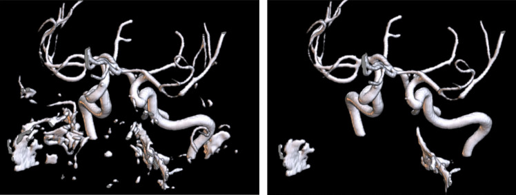
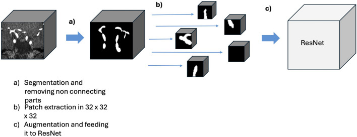
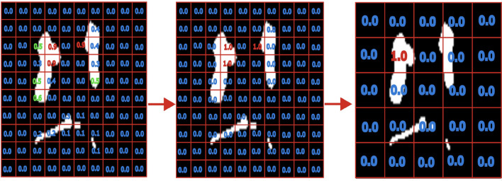
Intracranial atherosclerotic stenosis (ICAS) and intracranial aneurysms are prevalent conditions in the cerebrovascular system. ICAS causes a narrowing of the arterial lumen, thereby restricting blood flow, while aneurysms involve the ballooning of blood vessels. Both conditions can lead to severe outcomes, such as stroke or vessel rupture, which can be fatal. Early detection is crucial for effective intervention. In this study, we introduced a method that combines classical computer vision techniques with deep learning to detect intracranial aneurysms and ICAS in time-of-flight magnetic resonance angiography images. The process began with skull-stripping, followed by an affine transformation to align the images to a common atlas space. We then focused on the region of interest, including the circle of Willis, by cropping the relevant area. A segmentation algorithm was used to isolate the arteries, after which a patch-wise residual neural network was applied across the image. A voting mechanism was then employed to identify the presence of atrophies. Our method achieved accuracies of 76.5% for aneurysms and 82.4% for ICAS. Notably, when occlusions were not considered, the accuracy for ICAS detection improved to 85.7%. While the algorithm performed well for localized pathological findings, it was less effective at detecting occlusions, which involved long-range dependencies in the MRIs. This limitation was due to the architectural design of the patch-wise deep learning approach. Regardless, this can, in the future, be mitigated in a multi-scale patch-wise algorithm.
期刊介绍:
Neuroinformatics publishes original articles and reviews with an emphasis on data structure and software tools related to analysis, modeling, integration, and sharing in all areas of neuroscience research. The editors particularly invite contributions on: (1) Theory and methodology, including discussions on ontologies, modeling approaches, database design, and meta-analyses; (2) Descriptions of developed databases and software tools, and of the methods for their distribution; (3) Relevant experimental results, such as reports accompanie by the release of massive data sets; (4) Computational simulations of models integrating and organizing complex data; and (5) Neuroengineering approaches, including hardware, robotics, and information theory studies.


 分享
分享
 求助内容:
求助内容: 应助结果提醒方式:
 扫码关注我们
