{"title":"富生长因子血浆和含白细胞富血小板血浆对牙龈上皮细胞增殖和伤口愈合的影响。","authors":"Yuri Watanabe, Katsumitsu Shimada, Yousuke Doi, Takuyoshi Higuchi, Yoshiya Kato, Xianqi Li, Yuji Kurihara, Satoshi Murakami","doi":"10.1055/s-0044-1801274","DOIUrl":null,"url":null,"abstract":"<p><p>Plasma rich in growth factors (PRGF) is presumed to be able to stimulate the regeneration of skin and periodontal tissue. This effect can be attributed to the fact that PRGF contains fewer leukocyte-derived interleukins in comparison to platelet-rich plasma (PRP). However, a comparison of the effects of PRGF and PRP on gingival epithelial cells has not been conducted yet. Therefore, our objective was to clarify and compare the effects of PRGF and PRP on gingival epithelial cell proliferation, wound healing, and gene expression.PRGF and PRP were obtained from three donors. A complete medium containing bovine pituitary extract (BPE) and growth factors was used as a positive control (PC), while a medium without BPE was used as a negative control (NC). We evaluated the presence of platelets and leukocytes, as well as the number of leukocytes, in PRP and PRGF using the cell block method and a cell counting chamber. We assessed gingival epithelial cell proliferation with WST-1 and wound healing by using cell-free culture inserts. To examine the mRNA expression of tumor necrosis factor-α (TNF-α), which is related to cell growth inhibition, and integrin β4, which contributes to cell adhesion, we used quantitative reverse transcription polymerase chain reactions (RT-PCRs) under PRGF and PRP samples in vitro. The nonparametric data were analyzed using the Kruskal-Wallis test.Large quantities of platelets were observed in both PRGF and PRP. The leukocyte concentration in PRGF was generally lower than that in PRP. Our report indicated that cell proliferation was significantly higher in PRGF than in PRP on day 1 and 2. We found that there was no significant difference in the wound closure rate between PRGF and PRP in comparison to their respective control groups. The quantitative RT-PCR revealed insignificant differences in mRNA expression as TNF-α and integrin β4 between PRGF and PRP in comparison to the each of their respective control groups.Our research indicated that PRGF can promote the proliferation of gingival epithelium more than PRP, contributing to the healing of periodontal tissue. TNF-α and integrin β4 mRNA expression may not be significantly involved in wound closure within the gingival epithelium under the influence of PRGF and PRP.</p>","PeriodicalId":12028,"journal":{"name":"European Journal of Dentistry","volume":" ","pages":"1055-1062"},"PeriodicalIF":2.1000,"publicationDate":"2025-10-01","publicationTypes":"Journal Article","fieldsOfStudy":null,"isOpenAccess":false,"openAccessPdf":"https://www.ncbi.nlm.nih.gov/pmc/articles/PMC12494442/pdf/","citationCount":"0","resultStr":"{\"title\":\"A Comparative Analysis of Cell Proliferation and Wound Closure in Cultured Gingival Epithelial Cells Using Plasma Rich in Growth Factors and Platelet-Rich Plasma Containing Leukocytes.\",\"authors\":\"Yuri Watanabe, Katsumitsu Shimada, Yousuke Doi, Takuyoshi Higuchi, Yoshiya Kato, Xianqi Li, Yuji Kurihara, Satoshi Murakami\",\"doi\":\"10.1055/s-0044-1801274\",\"DOIUrl\":null,\"url\":null,\"abstract\":\"<p><p>Plasma rich in growth factors (PRGF) is presumed to be able to stimulate the regeneration of skin and periodontal tissue. This effect can be attributed to the fact that PRGF contains fewer leukocyte-derived interleukins in comparison to platelet-rich plasma (PRP). However, a comparison of the effects of PRGF and PRP on gingival epithelial cells has not been conducted yet. Therefore, our objective was to clarify and compare the effects of PRGF and PRP on gingival epithelial cell proliferation, wound healing, and gene expression.PRGF and PRP were obtained from three donors. A complete medium containing bovine pituitary extract (BPE) and growth factors was used as a positive control (PC), while a medium without BPE was used as a negative control (NC). We evaluated the presence of platelets and leukocytes, as well as the number of leukocytes, in PRP and PRGF using the cell block method and a cell counting chamber. We assessed gingival epithelial cell proliferation with WST-1 and wound healing by using cell-free culture inserts. To examine the mRNA expression of tumor necrosis factor-α (TNF-α), which is related to cell growth inhibition, and integrin β4, which contributes to cell adhesion, we used quantitative reverse transcription polymerase chain reactions (RT-PCRs) under PRGF and PRP samples in vitro. The nonparametric data were analyzed using the Kruskal-Wallis test.Large quantities of platelets were observed in both PRGF and PRP. The leukocyte concentration in PRGF was generally lower than that in PRP. Our report indicated that cell proliferation was significantly higher in PRGF than in PRP on day 1 and 2. We found that there was no significant difference in the wound closure rate between PRGF and PRP in comparison to their respective control groups. The quantitative RT-PCR revealed insignificant differences in mRNA expression as TNF-α and integrin β4 between PRGF and PRP in comparison to the each of their respective control groups.Our research indicated that PRGF can promote the proliferation of gingival epithelium more than PRP, contributing to the healing of periodontal tissue. TNF-α and integrin β4 mRNA expression may not be significantly involved in wound closure within the gingival epithelium under the influence of PRGF and PRP.</p>\",\"PeriodicalId\":12028,\"journal\":{\"name\":\"European Journal of Dentistry\",\"volume\":\" \",\"pages\":\"1055-1062\"},\"PeriodicalIF\":2.1000,\"publicationDate\":\"2025-10-01\",\"publicationTypes\":\"Journal Article\",\"fieldsOfStudy\":null,\"isOpenAccess\":false,\"openAccessPdf\":\"https://www.ncbi.nlm.nih.gov/pmc/articles/PMC12494442/pdf/\",\"citationCount\":\"0\",\"resultStr\":null,\"platform\":\"Semanticscholar\",\"paperid\":null,\"PeriodicalName\":\"European Journal of Dentistry\",\"FirstCategoryId\":\"1085\",\"ListUrlMain\":\"https://doi.org/10.1055/s-0044-1801274\",\"RegionNum\":0,\"RegionCategory\":null,\"ArticlePicture\":[],\"TitleCN\":null,\"AbstractTextCN\":null,\"PMCID\":null,\"EPubDate\":\"2025/1/20 0:00:00\",\"PubModel\":\"Epub\",\"JCR\":\"Q1\",\"JCRName\":\"Dentistry\",\"Score\":null,\"Total\":0}","platform":"Semanticscholar","paperid":null,"PeriodicalName":"European Journal of Dentistry","FirstCategoryId":"1085","ListUrlMain":"https://doi.org/10.1055/s-0044-1801274","RegionNum":0,"RegionCategory":null,"ArticlePicture":[],"TitleCN":null,"AbstractTextCN":null,"PMCID":null,"EPubDate":"2025/1/20 0:00:00","PubModel":"Epub","JCR":"Q1","JCRName":"Dentistry","Score":null,"Total":0}
A Comparative Analysis of Cell Proliferation and Wound Closure in Cultured Gingival Epithelial Cells Using Plasma Rich in Growth Factors and Platelet-Rich Plasma Containing Leukocytes.
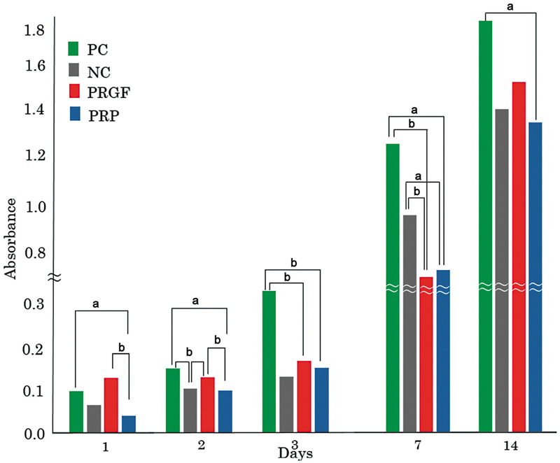
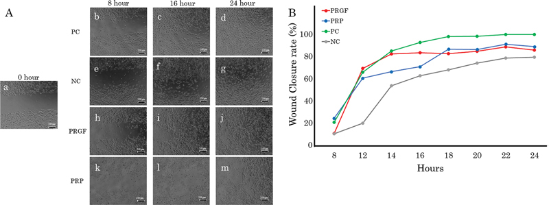
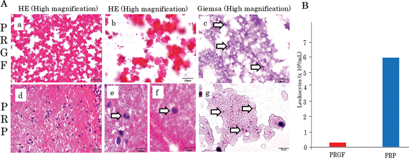
Plasma rich in growth factors (PRGF) is presumed to be able to stimulate the regeneration of skin and periodontal tissue. This effect can be attributed to the fact that PRGF contains fewer leukocyte-derived interleukins in comparison to platelet-rich plasma (PRP). However, a comparison of the effects of PRGF and PRP on gingival epithelial cells has not been conducted yet. Therefore, our objective was to clarify and compare the effects of PRGF and PRP on gingival epithelial cell proliferation, wound healing, and gene expression.PRGF and PRP were obtained from three donors. A complete medium containing bovine pituitary extract (BPE) and growth factors was used as a positive control (PC), while a medium without BPE was used as a negative control (NC). We evaluated the presence of platelets and leukocytes, as well as the number of leukocytes, in PRP and PRGF using the cell block method and a cell counting chamber. We assessed gingival epithelial cell proliferation with WST-1 and wound healing by using cell-free culture inserts. To examine the mRNA expression of tumor necrosis factor-α (TNF-α), which is related to cell growth inhibition, and integrin β4, which contributes to cell adhesion, we used quantitative reverse transcription polymerase chain reactions (RT-PCRs) under PRGF and PRP samples in vitro. The nonparametric data were analyzed using the Kruskal-Wallis test.Large quantities of platelets were observed in both PRGF and PRP. The leukocyte concentration in PRGF was generally lower than that in PRP. Our report indicated that cell proliferation was significantly higher in PRGF than in PRP on day 1 and 2. We found that there was no significant difference in the wound closure rate between PRGF and PRP in comparison to their respective control groups. The quantitative RT-PCR revealed insignificant differences in mRNA expression as TNF-α and integrin β4 between PRGF and PRP in comparison to the each of their respective control groups.Our research indicated that PRGF can promote the proliferation of gingival epithelium more than PRP, contributing to the healing of periodontal tissue. TNF-α and integrin β4 mRNA expression may not be significantly involved in wound closure within the gingival epithelium under the influence of PRGF and PRP.
期刊介绍:
The European Journal of Dentistry is the official journal of the Dental Investigations Society, based in Turkey. It is a double-blinded peer-reviewed, Open Access, multi-disciplinary international journal addressing various aspects of dentistry. The journal''s board consists of eminent investigators in dentistry from across the globe and presents an ideal international composition. The journal encourages its authors to submit original investigations, reviews, and reports addressing various divisions of dentistry including oral pathology, prosthodontics, endodontics, orthodontics etc. It is available both online and in print.


 分享
分享
 求助内容:
求助内容: 应助结果提醒方式:
 扫码关注我们
