Alberth Patricio Muñoz-Gualan, Abuzer Gungor, Monica Romano-Albornoz, Muhammet Enes Gurses, Cimen Elias, Arda Topcam, Serdar Ramanov, Ugur Ture
{"title":"小脑上-顶骨上入路:分离尸体大脑半球和保存中线结构的新方法。","authors":"Alberth Patricio Muñoz-Gualan, Abuzer Gungor, Monica Romano-Albornoz, Muhammet Enes Gurses, Cimen Elias, Arda Topcam, Serdar Ramanov, Ugur Ture","doi":"10.14744/SEMB.2024.92679","DOIUrl":null,"url":null,"abstract":"<p><strong>Objectives: </strong>To describe a novel technique for dissecting cadaver brains without damaging medial brain structures and surfaces, ensuring preservation for neuroanatomical study and training.</p><p><strong>Methods: </strong>Ten adult cadaveric brains were dissected using the supracerebellar suprapineal approach under an operative microscope with 6x to 40x magnification. This approach allowed for the separation of the brain into two hemispheres while providing direct visualization of the third ventricle and preserving midline structures.</p><p><strong>Results: </strong>The supracerebellar suprapineal approach enabled accurate and feasible dissection of the hemispheres without causing damage to the medial brain structures. All midline structures, including the third ventricle, were preserved, producing high-quality specimens for anatomical study.</p><p><strong>Conclusion: </strong>The supracerebellar suprapineal approach offers a significant advancement in the technique for hemispheric brain dissection, ensuring the preservation of medial brain structures and providing superior specimens for neurosurgical training and study.</p>","PeriodicalId":42218,"journal":{"name":"Medical Bulletin of Sisli Etfal Hospital","volume":"58 4","pages":"417-421"},"PeriodicalIF":1.0000,"publicationDate":"2024-12-24","publicationTypes":"Journal Article","fieldsOfStudy":null,"isOpenAccess":false,"openAccessPdf":"https://www.ncbi.nlm.nih.gov/pmc/articles/PMC11729829/pdf/","citationCount":"0","resultStr":"{\"title\":\"The Supracerebellar Suprapineal Approach: A Novel Method to Separate Cadaveric Brain Hemispheres and Preserve the Midline Structures.\",\"authors\":\"Alberth Patricio Muñoz-Gualan, Abuzer Gungor, Monica Romano-Albornoz, Muhammet Enes Gurses, Cimen Elias, Arda Topcam, Serdar Ramanov, Ugur Ture\",\"doi\":\"10.14744/SEMB.2024.92679\",\"DOIUrl\":null,\"url\":null,\"abstract\":\"<p><strong>Objectives: </strong>To describe a novel technique for dissecting cadaver brains without damaging medial brain structures and surfaces, ensuring preservation for neuroanatomical study and training.</p><p><strong>Methods: </strong>Ten adult cadaveric brains were dissected using the supracerebellar suprapineal approach under an operative microscope with 6x to 40x magnification. This approach allowed for the separation of the brain into two hemispheres while providing direct visualization of the third ventricle and preserving midline structures.</p><p><strong>Results: </strong>The supracerebellar suprapineal approach enabled accurate and feasible dissection of the hemispheres without causing damage to the medial brain structures. All midline structures, including the third ventricle, were preserved, producing high-quality specimens for anatomical study.</p><p><strong>Conclusion: </strong>The supracerebellar suprapineal approach offers a significant advancement in the technique for hemispheric brain dissection, ensuring the preservation of medial brain structures and providing superior specimens for neurosurgical training and study.</p>\",\"PeriodicalId\":42218,\"journal\":{\"name\":\"Medical Bulletin of Sisli Etfal Hospital\",\"volume\":\"58 4\",\"pages\":\"417-421\"},\"PeriodicalIF\":1.0000,\"publicationDate\":\"2024-12-24\",\"publicationTypes\":\"Journal Article\",\"fieldsOfStudy\":null,\"isOpenAccess\":false,\"openAccessPdf\":\"https://www.ncbi.nlm.nih.gov/pmc/articles/PMC11729829/pdf/\",\"citationCount\":\"0\",\"resultStr\":null,\"platform\":\"Semanticscholar\",\"paperid\":null,\"PeriodicalName\":\"Medical Bulletin of Sisli Etfal Hospital\",\"FirstCategoryId\":\"1085\",\"ListUrlMain\":\"https://doi.org/10.14744/SEMB.2024.92679\",\"RegionNum\":0,\"RegionCategory\":null,\"ArticlePicture\":[],\"TitleCN\":null,\"AbstractTextCN\":null,\"PMCID\":null,\"EPubDate\":\"2024/1/1 0:00:00\",\"PubModel\":\"eCollection\",\"JCR\":\"Q3\",\"JCRName\":\"MEDICINE, GENERAL & INTERNAL\",\"Score\":null,\"Total\":0}","platform":"Semanticscholar","paperid":null,"PeriodicalName":"Medical Bulletin of Sisli Etfal Hospital","FirstCategoryId":"1085","ListUrlMain":"https://doi.org/10.14744/SEMB.2024.92679","RegionNum":0,"RegionCategory":null,"ArticlePicture":[],"TitleCN":null,"AbstractTextCN":null,"PMCID":null,"EPubDate":"2024/1/1 0:00:00","PubModel":"eCollection","JCR":"Q3","JCRName":"MEDICINE, GENERAL & INTERNAL","Score":null,"Total":0}
The Supracerebellar Suprapineal Approach: A Novel Method to Separate Cadaveric Brain Hemispheres and Preserve the Midline Structures.
Objectives: To describe a novel technique for dissecting cadaver brains without damaging medial brain structures and surfaces, ensuring preservation for neuroanatomical study and training.
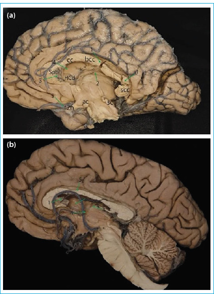
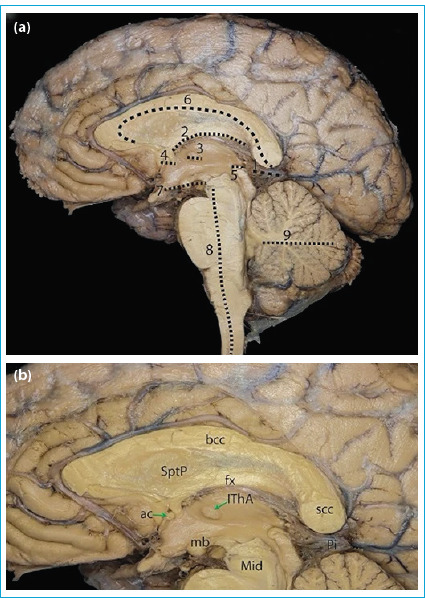
Methods: Ten adult cadaveric brains were dissected using the supracerebellar suprapineal approach under an operative microscope with 6x to 40x magnification. This approach allowed for the separation of the brain into two hemispheres while providing direct visualization of the third ventricle and preserving midline structures.
Results: The supracerebellar suprapineal approach enabled accurate and feasible dissection of the hemispheres without causing damage to the medial brain structures. All midline structures, including the third ventricle, were preserved, producing high-quality specimens for anatomical study.
Conclusion: The supracerebellar suprapineal approach offers a significant advancement in the technique for hemispheric brain dissection, ensuring the preservation of medial brain structures and providing superior specimens for neurosurgical training and study.


 分享
分享
 求助内容:
求助内容: 应助结果提醒方式:
 扫码关注我们
