{"title":"病理组织证实1例由疣状毛癣引起的角质细胞,伴有类似药疹的皮肤癣反应和粘膜感染。","authors":"Makoto Ishiai, Hiroshi Tanabe","doi":"10.1016/j.mmcr.2024.100691","DOIUrl":null,"url":null,"abstract":"<div><div>We report a case of kerion celsi caused by <em>Trichophyton tonsurans</em> in a teenage male judo athlete, presenting with a lesion in the occipital region. Following the initiation of systemic antifungal therapy, the patient developed a dermatophytid reaction, necessitating differentiation from a drug eruption. Direct microscopy of the affected area confirmed the presence of fungal elements, and histopathological examination revealed endothrix invasion, supporting the continuation of treatment. A drug-induced lymphocyte stimulation test for terbinafine, conducted post-treatment, was negative. This case highlights the importance of distinguishing dermatophytid reactions from drug eruptions to ensure uninterrupted antifungal therapy.</div></div>","PeriodicalId":51724,"journal":{"name":"Medical Mycology Case Reports","volume":"47 ","pages":"Article 100691"},"PeriodicalIF":1.3000,"publicationDate":"2025-03-01","publicationTypes":"Journal Article","fieldsOfStudy":null,"isOpenAccess":false,"openAccessPdf":"https://www.ncbi.nlm.nih.gov/pmc/articles/PMC11750295/pdf/","citationCount":"0","resultStr":"{\"title\":\"A case of kerion celsi caused by Trichophyton tonsurans with dermatophytid reaction mimicking a drug eruption and endothrix infection confirmed in pathological tissue\",\"authors\":\"Makoto Ishiai, Hiroshi Tanabe\",\"doi\":\"10.1016/j.mmcr.2024.100691\",\"DOIUrl\":null,\"url\":null,\"abstract\":\"<div><div>We report a case of kerion celsi caused by <em>Trichophyton tonsurans</em> in a teenage male judo athlete, presenting with a lesion in the occipital region. Following the initiation of systemic antifungal therapy, the patient developed a dermatophytid reaction, necessitating differentiation from a drug eruption. Direct microscopy of the affected area confirmed the presence of fungal elements, and histopathological examination revealed endothrix invasion, supporting the continuation of treatment. A drug-induced lymphocyte stimulation test for terbinafine, conducted post-treatment, was negative. This case highlights the importance of distinguishing dermatophytid reactions from drug eruptions to ensure uninterrupted antifungal therapy.</div></div>\",\"PeriodicalId\":51724,\"journal\":{\"name\":\"Medical Mycology Case Reports\",\"volume\":\"47 \",\"pages\":\"Article 100691\"},\"PeriodicalIF\":1.3000,\"publicationDate\":\"2025-03-01\",\"publicationTypes\":\"Journal Article\",\"fieldsOfStudy\":null,\"isOpenAccess\":false,\"openAccessPdf\":\"https://www.ncbi.nlm.nih.gov/pmc/articles/PMC11750295/pdf/\",\"citationCount\":\"0\",\"resultStr\":null,\"platform\":\"Semanticscholar\",\"paperid\":null,\"PeriodicalName\":\"Medical Mycology Case Reports\",\"FirstCategoryId\":\"1085\",\"ListUrlMain\":\"https://www.sciencedirect.com/science/article/pii/S2211753924000654\",\"RegionNum\":0,\"RegionCategory\":null,\"ArticlePicture\":[],\"TitleCN\":null,\"AbstractTextCN\":null,\"PMCID\":null,\"EPubDate\":\"2024/12/25 0:00:00\",\"PubModel\":\"Epub\",\"JCR\":\"Q3\",\"JCRName\":\"MEDICINE, RESEARCH & EXPERIMENTAL\",\"Score\":null,\"Total\":0}","platform":"Semanticscholar","paperid":null,"PeriodicalName":"Medical Mycology Case Reports","FirstCategoryId":"1085","ListUrlMain":"https://www.sciencedirect.com/science/article/pii/S2211753924000654","RegionNum":0,"RegionCategory":null,"ArticlePicture":[],"TitleCN":null,"AbstractTextCN":null,"PMCID":null,"EPubDate":"2024/12/25 0:00:00","PubModel":"Epub","JCR":"Q3","JCRName":"MEDICINE, RESEARCH & EXPERIMENTAL","Score":null,"Total":0}
A case of kerion celsi caused by Trichophyton tonsurans with dermatophytid reaction mimicking a drug eruption and endothrix infection confirmed in pathological tissue
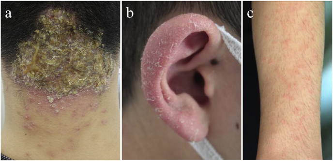
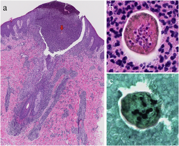
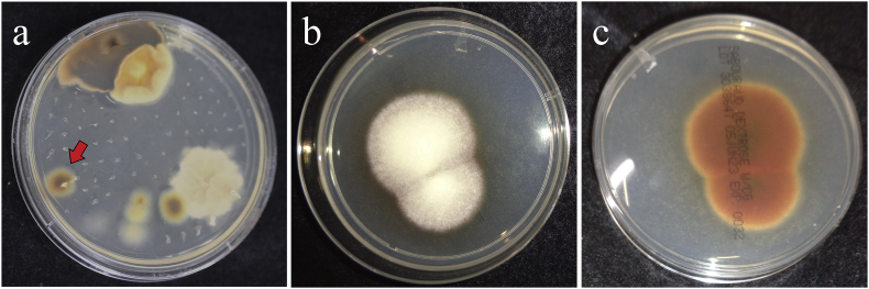
We report a case of kerion celsi caused by Trichophyton tonsurans in a teenage male judo athlete, presenting with a lesion in the occipital region. Following the initiation of systemic antifungal therapy, the patient developed a dermatophytid reaction, necessitating differentiation from a drug eruption. Direct microscopy of the affected area confirmed the presence of fungal elements, and histopathological examination revealed endothrix invasion, supporting the continuation of treatment. A drug-induced lymphocyte stimulation test for terbinafine, conducted post-treatment, was negative. This case highlights the importance of distinguishing dermatophytid reactions from drug eruptions to ensure uninterrupted antifungal therapy.


 分享
分享
 求助内容:
求助内容: 应助结果提醒方式:
 扫码关注我们
