Trine Amalie Fogh Gadeberg, Martin Høgholm Jørgensen, Heidi Gytz Olesen, Josefine Lorentzen, Seandean Lykke Harwood, Ana Viana Almeida, Marlene Uglebjerg Fruergaard, Rasmus Kjeldsen Jensen, Philipp Kanis, Henrik Pedersen, Emil Tranchant, Steen Vang Petersen, Ida Buch Thøgersen, Birthe Brandt Kragelund, Joseph Anthony Lyons, Jan Johannes Enghild, Gregers Rom Andersen
{"title":"补体C3的低温电镜分析显示巨球蛋白环有一个可逆的大开口","authors":"Trine Amalie Fogh Gadeberg, Martin Høgholm Jørgensen, Heidi Gytz Olesen, Josefine Lorentzen, Seandean Lykke Harwood, Ana Viana Almeida, Marlene Uglebjerg Fruergaard, Rasmus Kjeldsen Jensen, Philipp Kanis, Henrik Pedersen, Emil Tranchant, Steen Vang Petersen, Ida Buch Thøgersen, Birthe Brandt Kragelund, Joseph Anthony Lyons, Jan Johannes Enghild, Gregers Rom Andersen","doi":"10.1038/s41594-024-01467-4","DOIUrl":null,"url":null,"abstract":"The C3 protein is the central molecule within the complement system and undergoes proteolytic activation to C3b in the presence of pathogens. Pattern-independent activation of C3 also occurs via hydrolysis, resulting in C3(H2O), but the structural details of C3 hydrolysis remain elusive. Here we show that the conformation of the C3(H2O) analog, C3MA, is indistinguishable from C3b. In contrast, the reaction intermediate C3* adopts a conformation dramatically different from both C3 and C3MA. In C3*, unlocking of the macroglobulin (MG) 3 domain creates a large opening in the MG ring through which the anaphylatoxin (ANA) domain translocates through a transient opening. C3MA formation is inhibited by an MG3-specific nanobody and prevented by linking the ANA domain to the C3 β-chain. Our study reveals an unexpected dynamic behavior of C3 and forms the basis for elucidation of the in vivo contribution of C3 hydrolysis and for controlling complement upon intravascular hemolysis and surface-contact-induced activation. The structure of a spontaneously activated immune protein is determined by cryo-electron microscopy. This reveals the passage of an entire domain through a transient opening. The authors investigate the mechanism by multiple biophysical approaches.","PeriodicalId":49141,"journal":{"name":"Nature Structural & Molecular Biology","volume":"32 5","pages":"884-895"},"PeriodicalIF":10.1000,"publicationDate":"2025-01-23","publicationTypes":"Journal Article","fieldsOfStudy":null,"isOpenAccess":false,"openAccessPdf":"","citationCount":"0","resultStr":"{\"title\":\"Cryo-EM analysis of complement C3 reveals a reversible major opening of the macroglobulin ring\",\"authors\":\"Trine Amalie Fogh Gadeberg, Martin Høgholm Jørgensen, Heidi Gytz Olesen, Josefine Lorentzen, Seandean Lykke Harwood, Ana Viana Almeida, Marlene Uglebjerg Fruergaard, Rasmus Kjeldsen Jensen, Philipp Kanis, Henrik Pedersen, Emil Tranchant, Steen Vang Petersen, Ida Buch Thøgersen, Birthe Brandt Kragelund, Joseph Anthony Lyons, Jan Johannes Enghild, Gregers Rom Andersen\",\"doi\":\"10.1038/s41594-024-01467-4\",\"DOIUrl\":null,\"url\":null,\"abstract\":\"The C3 protein is the central molecule within the complement system and undergoes proteolytic activation to C3b in the presence of pathogens. Pattern-independent activation of C3 also occurs via hydrolysis, resulting in C3(H2O), but the structural details of C3 hydrolysis remain elusive. Here we show that the conformation of the C3(H2O) analog, C3MA, is indistinguishable from C3b. In contrast, the reaction intermediate C3* adopts a conformation dramatically different from both C3 and C3MA. In C3*, unlocking of the macroglobulin (MG) 3 domain creates a large opening in the MG ring through which the anaphylatoxin (ANA) domain translocates through a transient opening. C3MA formation is inhibited by an MG3-specific nanobody and prevented by linking the ANA domain to the C3 β-chain. Our study reveals an unexpected dynamic behavior of C3 and forms the basis for elucidation of the in vivo contribution of C3 hydrolysis and for controlling complement upon intravascular hemolysis and surface-contact-induced activation. The structure of a spontaneously activated immune protein is determined by cryo-electron microscopy. This reveals the passage of an entire domain through a transient opening. The authors investigate the mechanism by multiple biophysical approaches.\",\"PeriodicalId\":49141,\"journal\":{\"name\":\"Nature Structural & Molecular Biology\",\"volume\":\"32 5\",\"pages\":\"884-895\"},\"PeriodicalIF\":10.1000,\"publicationDate\":\"2025-01-23\",\"publicationTypes\":\"Journal Article\",\"fieldsOfStudy\":null,\"isOpenAccess\":false,\"openAccessPdf\":\"\",\"citationCount\":\"0\",\"resultStr\":null,\"platform\":\"Semanticscholar\",\"paperid\":null,\"PeriodicalName\":\"Nature Structural & Molecular Biology\",\"FirstCategoryId\":\"99\",\"ListUrlMain\":\"https://www.nature.com/articles/s41594-024-01467-4\",\"RegionNum\":1,\"RegionCategory\":\"生物学\",\"ArticlePicture\":[],\"TitleCN\":null,\"AbstractTextCN\":null,\"PMCID\":null,\"EPubDate\":\"\",\"PubModel\":\"\",\"JCR\":\"Q1\",\"JCRName\":\"BIOCHEMISTRY & MOLECULAR BIOLOGY\",\"Score\":null,\"Total\":0}","platform":"Semanticscholar","paperid":null,"PeriodicalName":"Nature Structural & Molecular Biology","FirstCategoryId":"99","ListUrlMain":"https://www.nature.com/articles/s41594-024-01467-4","RegionNum":1,"RegionCategory":"生物学","ArticlePicture":[],"TitleCN":null,"AbstractTextCN":null,"PMCID":null,"EPubDate":"","PubModel":"","JCR":"Q1","JCRName":"BIOCHEMISTRY & MOLECULAR BIOLOGY","Score":null,"Total":0}
Cryo-EM analysis of complement C3 reveals a reversible major opening of the macroglobulin ring
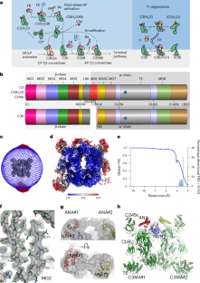
The C3 protein is the central molecule within the complement system and undergoes proteolytic activation to C3b in the presence of pathogens. Pattern-independent activation of C3 also occurs via hydrolysis, resulting in C3(H2O), but the structural details of C3 hydrolysis remain elusive. Here we show that the conformation of the C3(H2O) analog, C3MA, is indistinguishable from C3b. In contrast, the reaction intermediate C3* adopts a conformation dramatically different from both C3 and C3MA. In C3*, unlocking of the macroglobulin (MG) 3 domain creates a large opening in the MG ring through which the anaphylatoxin (ANA) domain translocates through a transient opening. C3MA formation is inhibited by an MG3-specific nanobody and prevented by linking the ANA domain to the C3 β-chain. Our study reveals an unexpected dynamic behavior of C3 and forms the basis for elucidation of the in vivo contribution of C3 hydrolysis and for controlling complement upon intravascular hemolysis and surface-contact-induced activation. The structure of a spontaneously activated immune protein is determined by cryo-electron microscopy. This reveals the passage of an entire domain through a transient opening. The authors investigate the mechanism by multiple biophysical approaches.
期刊介绍:
Nature Structural & Molecular Biology is a comprehensive platform that combines structural and molecular research. Our journal focuses on exploring the functional and mechanistic aspects of biological processes, emphasizing how molecular components collaborate to achieve a particular function. While structural data can shed light on these insights, our publication does not require them as a prerequisite.


 分享
分享
 求助内容:
求助内容: 应助结果提醒方式:
 扫码关注我们
