{"title":"GenAI 从拉曼成像合成组织病理学图像,用于术中舌鳞状细胞癌评估","authors":"Bing Yan, Zhining Wen, Lili Xue, Tianyi Wang, Zhichao Liu, Wulin Long, Yi Li, Runyu Jing","doi":"10.1038/s41368-025-00346-y","DOIUrl":null,"url":null,"abstract":"<p>The presence of a positive deep surgical margin in tongue squamous cell carcinoma (TSCC) significantly elevates the risk of local recurrence. Therefore, a prompt and precise intraoperative assessment of margin status is imperative to ensure thorough tumor resection. In this study, we integrate Raman imaging technology with an artificial intelligence (AI) generative model, proposing an innovative approach for intraoperative margin status diagnosis. This method utilizes Raman imaging to swiftly and non-invasively capture tissue Raman images, which are then transformed into hematoxylin-eosin (H&E)-stained histopathological images using an AI generative model for histopathological diagnosis. The generated H&E-stained images clearly illustrate the tissue’s pathological conditions. Independently reviewed by three pathologists, the overall diagnostic accuracy for distinguishing between tumor tissue and normal muscle tissue reaches 86.7%. Notably, it outperforms current clinical practices, especially in TSCC with positive lymph node metastasis or moderately differentiated grades. This advancement highlights the potential of AI-enhanced Raman imaging to significantly improve intraoperative assessments and surgical margin evaluations, promising a versatile diagnostic tool beyond TSCC.</p>","PeriodicalId":14191,"journal":{"name":"International Journal of Oral Science","volume":"45 1","pages":""},"PeriodicalIF":12.2000,"publicationDate":"2025-01-26","publicationTypes":"Journal Article","fieldsOfStudy":null,"isOpenAccess":false,"openAccessPdf":"","citationCount":"0","resultStr":"{\"title\":\"GenAI synthesis of histopathological images from Raman imaging for intraoperative tongue squamous cell carcinoma assessment\",\"authors\":\"Bing Yan, Zhining Wen, Lili Xue, Tianyi Wang, Zhichao Liu, Wulin Long, Yi Li, Runyu Jing\",\"doi\":\"10.1038/s41368-025-00346-y\",\"DOIUrl\":null,\"url\":null,\"abstract\":\"<p>The presence of a positive deep surgical margin in tongue squamous cell carcinoma (TSCC) significantly elevates the risk of local recurrence. Therefore, a prompt and precise intraoperative assessment of margin status is imperative to ensure thorough tumor resection. In this study, we integrate Raman imaging technology with an artificial intelligence (AI) generative model, proposing an innovative approach for intraoperative margin status diagnosis. This method utilizes Raman imaging to swiftly and non-invasively capture tissue Raman images, which are then transformed into hematoxylin-eosin (H&E)-stained histopathological images using an AI generative model for histopathological diagnosis. The generated H&E-stained images clearly illustrate the tissue’s pathological conditions. Independently reviewed by three pathologists, the overall diagnostic accuracy for distinguishing between tumor tissue and normal muscle tissue reaches 86.7%. Notably, it outperforms current clinical practices, especially in TSCC with positive lymph node metastasis or moderately differentiated grades. This advancement highlights the potential of AI-enhanced Raman imaging to significantly improve intraoperative assessments and surgical margin evaluations, promising a versatile diagnostic tool beyond TSCC.</p>\",\"PeriodicalId\":14191,\"journal\":{\"name\":\"International Journal of Oral Science\",\"volume\":\"45 1\",\"pages\":\"\"},\"PeriodicalIF\":12.2000,\"publicationDate\":\"2025-01-26\",\"publicationTypes\":\"Journal Article\",\"fieldsOfStudy\":null,\"isOpenAccess\":false,\"openAccessPdf\":\"\",\"citationCount\":\"0\",\"resultStr\":null,\"platform\":\"Semanticscholar\",\"paperid\":null,\"PeriodicalName\":\"International Journal of Oral Science\",\"FirstCategoryId\":\"3\",\"ListUrlMain\":\"https://doi.org/10.1038/s41368-025-00346-y\",\"RegionNum\":1,\"RegionCategory\":\"医学\",\"ArticlePicture\":[],\"TitleCN\":null,\"AbstractTextCN\":null,\"PMCID\":null,\"EPubDate\":\"\",\"PubModel\":\"\",\"JCR\":\"Q1\",\"JCRName\":\"DENTISTRY, ORAL SURGERY & MEDICINE\",\"Score\":null,\"Total\":0}","platform":"Semanticscholar","paperid":null,"PeriodicalName":"International Journal of Oral Science","FirstCategoryId":"3","ListUrlMain":"https://doi.org/10.1038/s41368-025-00346-y","RegionNum":1,"RegionCategory":"医学","ArticlePicture":[],"TitleCN":null,"AbstractTextCN":null,"PMCID":null,"EPubDate":"","PubModel":"","JCR":"Q1","JCRName":"DENTISTRY, ORAL SURGERY & MEDICINE","Score":null,"Total":0}
GenAI synthesis of histopathological images from Raman imaging for intraoperative tongue squamous cell carcinoma assessment
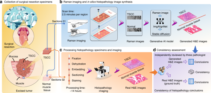
The presence of a positive deep surgical margin in tongue squamous cell carcinoma (TSCC) significantly elevates the risk of local recurrence. Therefore, a prompt and precise intraoperative assessment of margin status is imperative to ensure thorough tumor resection. In this study, we integrate Raman imaging technology with an artificial intelligence (AI) generative model, proposing an innovative approach for intraoperative margin status diagnosis. This method utilizes Raman imaging to swiftly and non-invasively capture tissue Raman images, which are then transformed into hematoxylin-eosin (H&E)-stained histopathological images using an AI generative model for histopathological diagnosis. The generated H&E-stained images clearly illustrate the tissue’s pathological conditions. Independently reviewed by three pathologists, the overall diagnostic accuracy for distinguishing between tumor tissue and normal muscle tissue reaches 86.7%. Notably, it outperforms current clinical practices, especially in TSCC with positive lymph node metastasis or moderately differentiated grades. This advancement highlights the potential of AI-enhanced Raman imaging to significantly improve intraoperative assessments and surgical margin evaluations, promising a versatile diagnostic tool beyond TSCC.
期刊介绍:
The International Journal of Oral Science covers various aspects of oral science and interdisciplinary fields, encompassing basic, applied, and clinical research. Topics include, but are not limited to:
Oral microbiology
Oral and maxillofacial oncology
Cariology
Oral inflammation and infection
Dental stem cells and regenerative medicine
Craniofacial surgery
Dental material
Oral biomechanics
Oral, dental, and maxillofacial genetic and developmental diseases
Craniofacial bone research
Craniofacial-related biomaterials
Temporomandibular joint disorder and osteoarthritis
The journal publishes peer-reviewed Articles presenting new research results and Review Articles offering concise summaries of specific areas in oral science.


 分享
分享
 求助内容:
求助内容: 应助结果提醒方式:
 扫码关注我们
