Abdul Al-Shawwa, Michael Craig, Kalum Ost, David Anderson, Steve Casha, W Bradley Jacobs, Nathan Evaniew, Saswati Tripathy, Jacques Bouchard, Peter Lewkonia, Fred Nicholls, Alex Soroceanu, Ganesh Swamy, Kenneth C Thomas, Stephan duPlessis, Michael Mh Yang, Julien Cohen-Adad, Nicholas Dea, Jefferson R Wilson, David W Cadotte
{"title":"脊髓脱髓鞘可预测轻度退行性颈椎病患者的神经功能衰退。","authors":"Abdul Al-Shawwa, Michael Craig, Kalum Ost, David Anderson, Steve Casha, W Bradley Jacobs, Nathan Evaniew, Saswati Tripathy, Jacques Bouchard, Peter Lewkonia, Fred Nicholls, Alex Soroceanu, Ganesh Swamy, Kenneth C Thomas, Stephan duPlessis, Michael Mh Yang, Julien Cohen-Adad, Nicholas Dea, Jefferson R Wilson, David W Cadotte","doi":"10.1136/bmjno-2024-000940","DOIUrl":null,"url":null,"abstract":"<p><strong>Background: </strong>Degenerative cervical myelopathy (DCM) is the most common form of atraumatic spinal cord injury globally. Clinical guidelines regarding surgery for patients with mild DCM and minimal symptoms remain uncertain. This study aims to identify imaging and clinical predictors of neurological deterioration in mild DCM and explore pathophysiological correlates to guide clinical decision-making.</p><p><strong>Methods: </strong>Patients with mild DCM underwent advanced MRI scans that included T2-weighted, diffusion tensor imaging and magnetisation transfer (MT) sequences, along with clinical outcome measures at baseline and 6-month intervals after enrolment. Quantitative MRI (qMRI) metrics were derived above and below maximally compressed cervical levels (MCCLs). Various machine learning (ML) models were trained to predict 6 month neurological deterioration, followed by global and local model interpretation to assess feature importance.</p><p><strong>Results: </strong>A total of 49 patients were followed for a maximum of 2 years, contributing 110 6-month data entries. Neurological deterioration occurred in 38% of cases. The best-performing ML model, combining clinical and qMRI metrics, achieved a balanced accuracy of 83%, and an area under curve-receiver operating characteristic of 0.87. Key predictors included MT ratio (demyelination) above the MCCL in the dorsal and ventral funiculi and moderate tingling in the arm, shoulder or hand. qMRI metrics significantly improved predictive performance compared to models using only clinical (bal. acc=68.1%) or imaging data (bal. acc=57.4%).</p><p><strong>Conclusions: </strong>Reduced myelin content in the dorsal and ventral funiculi above the site of compression, combined with sensory deficits in the hands and gait/balance disturbances, predicts 6-month neurological deterioration in mild DCM and may warrant early surgical intervention.</p>","PeriodicalId":52754,"journal":{"name":"BMJ Neurology Open","volume":"7 1","pages":"e000940"},"PeriodicalIF":2.4000,"publicationDate":"2025-01-31","publicationTypes":"Journal Article","fieldsOfStudy":null,"isOpenAccess":false,"openAccessPdf":"https://www.ncbi.nlm.nih.gov/pmc/articles/PMC11792293/pdf/","citationCount":"0","resultStr":"{\"title\":\"Spinal cord demyelination predicts neurological deterioration in patients with mild degenerative cervical myelopathy.\",\"authors\":\"Abdul Al-Shawwa, Michael Craig, Kalum Ost, David Anderson, Steve Casha, W Bradley Jacobs, Nathan Evaniew, Saswati Tripathy, Jacques Bouchard, Peter Lewkonia, Fred Nicholls, Alex Soroceanu, Ganesh Swamy, Kenneth C Thomas, Stephan duPlessis, Michael Mh Yang, Julien Cohen-Adad, Nicholas Dea, Jefferson R Wilson, David W Cadotte\",\"doi\":\"10.1136/bmjno-2024-000940\",\"DOIUrl\":null,\"url\":null,\"abstract\":\"<p><strong>Background: </strong>Degenerative cervical myelopathy (DCM) is the most common form of atraumatic spinal cord injury globally. Clinical guidelines regarding surgery for patients with mild DCM and minimal symptoms remain uncertain. This study aims to identify imaging and clinical predictors of neurological deterioration in mild DCM and explore pathophysiological correlates to guide clinical decision-making.</p><p><strong>Methods: </strong>Patients with mild DCM underwent advanced MRI scans that included T2-weighted, diffusion tensor imaging and magnetisation transfer (MT) sequences, along with clinical outcome measures at baseline and 6-month intervals after enrolment. Quantitative MRI (qMRI) metrics were derived above and below maximally compressed cervical levels (MCCLs). Various machine learning (ML) models were trained to predict 6 month neurological deterioration, followed by global and local model interpretation to assess feature importance.</p><p><strong>Results: </strong>A total of 49 patients were followed for a maximum of 2 years, contributing 110 6-month data entries. Neurological deterioration occurred in 38% of cases. The best-performing ML model, combining clinical and qMRI metrics, achieved a balanced accuracy of 83%, and an area under curve-receiver operating characteristic of 0.87. Key predictors included MT ratio (demyelination) above the MCCL in the dorsal and ventral funiculi and moderate tingling in the arm, shoulder or hand. qMRI metrics significantly improved predictive performance compared to models using only clinical (bal. acc=68.1%) or imaging data (bal. acc=57.4%).</p><p><strong>Conclusions: </strong>Reduced myelin content in the dorsal and ventral funiculi above the site of compression, combined with sensory deficits in the hands and gait/balance disturbances, predicts 6-month neurological deterioration in mild DCM and may warrant early surgical intervention.</p>\",\"PeriodicalId\":52754,\"journal\":{\"name\":\"BMJ Neurology Open\",\"volume\":\"7 1\",\"pages\":\"e000940\"},\"PeriodicalIF\":2.4000,\"publicationDate\":\"2025-01-31\",\"publicationTypes\":\"Journal Article\",\"fieldsOfStudy\":null,\"isOpenAccess\":false,\"openAccessPdf\":\"https://www.ncbi.nlm.nih.gov/pmc/articles/PMC11792293/pdf/\",\"citationCount\":\"0\",\"resultStr\":null,\"platform\":\"Semanticscholar\",\"paperid\":null,\"PeriodicalName\":\"BMJ Neurology Open\",\"FirstCategoryId\":\"1085\",\"ListUrlMain\":\"https://doi.org/10.1136/bmjno-2024-000940\",\"RegionNum\":0,\"RegionCategory\":null,\"ArticlePicture\":[],\"TitleCN\":null,\"AbstractTextCN\":null,\"PMCID\":null,\"EPubDate\":\"2025/1/1 0:00:00\",\"PubModel\":\"eCollection\",\"JCR\":\"Q3\",\"JCRName\":\"CLINICAL NEUROLOGY\",\"Score\":null,\"Total\":0}","platform":"Semanticscholar","paperid":null,"PeriodicalName":"BMJ Neurology Open","FirstCategoryId":"1085","ListUrlMain":"https://doi.org/10.1136/bmjno-2024-000940","RegionNum":0,"RegionCategory":null,"ArticlePicture":[],"TitleCN":null,"AbstractTextCN":null,"PMCID":null,"EPubDate":"2025/1/1 0:00:00","PubModel":"eCollection","JCR":"Q3","JCRName":"CLINICAL NEUROLOGY","Score":null,"Total":0}
Spinal cord demyelination predicts neurological deterioration in patients with mild degenerative cervical myelopathy.
Background: Degenerative cervical myelopathy (DCM) is the most common form of atraumatic spinal cord injury globally. Clinical guidelines regarding surgery for patients with mild DCM and minimal symptoms remain uncertain. This study aims to identify imaging and clinical predictors of neurological deterioration in mild DCM and explore pathophysiological correlates to guide clinical decision-making.
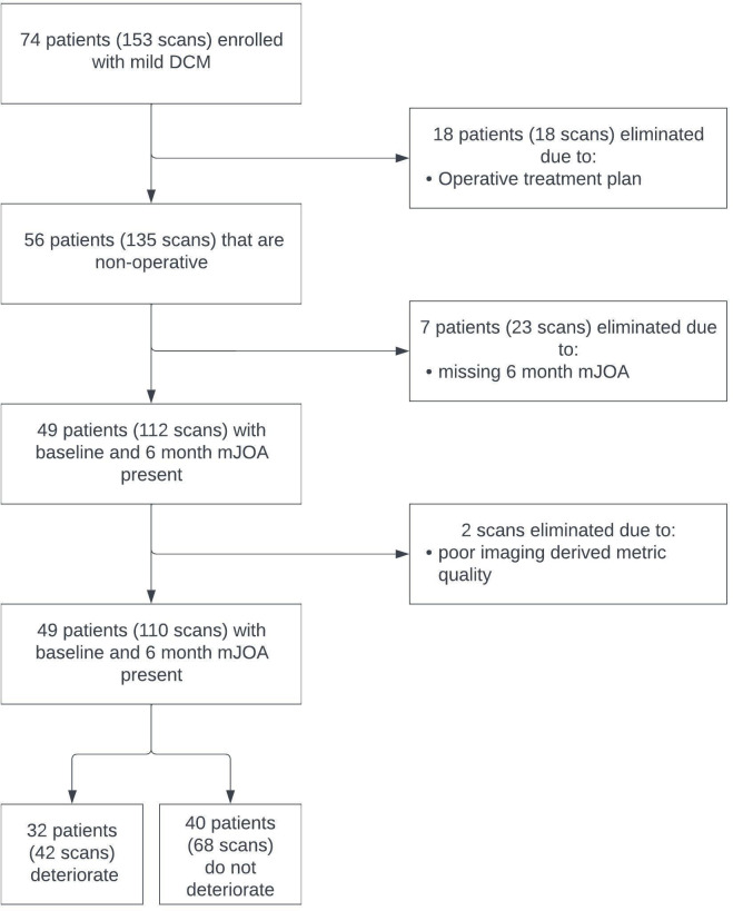
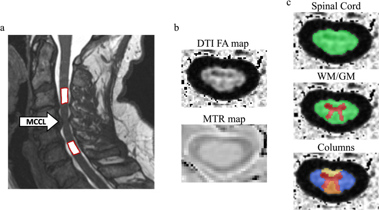
Methods: Patients with mild DCM underwent advanced MRI scans that included T2-weighted, diffusion tensor imaging and magnetisation transfer (MT) sequences, along with clinical outcome measures at baseline and 6-month intervals after enrolment. Quantitative MRI (qMRI) metrics were derived above and below maximally compressed cervical levels (MCCLs). Various machine learning (ML) models were trained to predict 6 month neurological deterioration, followed by global and local model interpretation to assess feature importance.
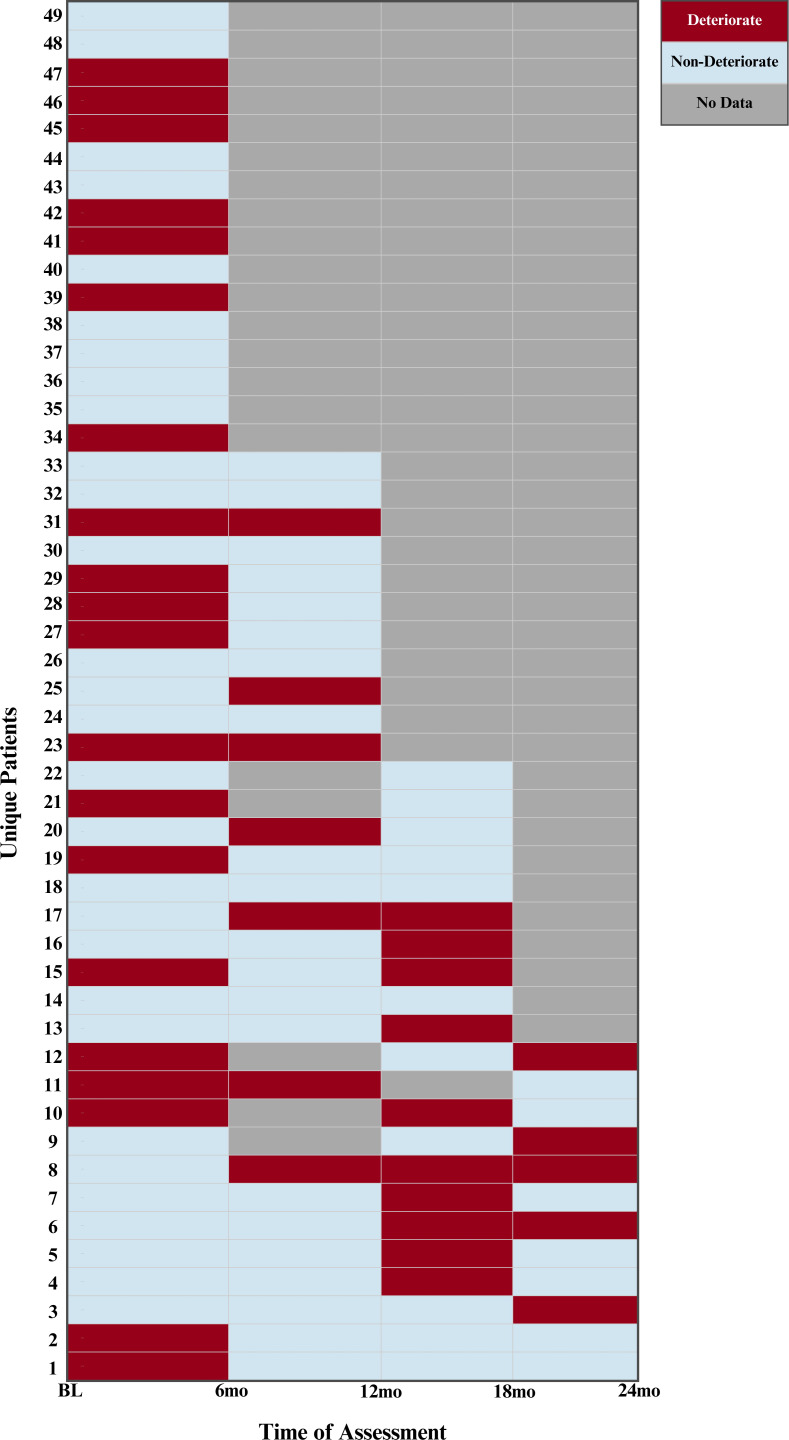
Results: A total of 49 patients were followed for a maximum of 2 years, contributing 110 6-month data entries. Neurological deterioration occurred in 38% of cases. The best-performing ML model, combining clinical and qMRI metrics, achieved a balanced accuracy of 83%, and an area under curve-receiver operating characteristic of 0.87. Key predictors included MT ratio (demyelination) above the MCCL in the dorsal and ventral funiculi and moderate tingling in the arm, shoulder or hand. qMRI metrics significantly improved predictive performance compared to models using only clinical (bal. acc=68.1%) or imaging data (bal. acc=57.4%).
Conclusions: Reduced myelin content in the dorsal and ventral funiculi above the site of compression, combined with sensory deficits in the hands and gait/balance disturbances, predicts 6-month neurological deterioration in mild DCM and may warrant early surgical intervention.


 分享
分享
 求助内容:
求助内容: 应助结果提醒方式:
 扫码关注我们
