{"title":"反射共聚焦显微镜对白癜风不同病变区域的敏感性和特异性分析","authors":"Jiyuan Wu, Miaomiao Sun, Zijia Wei, Qian Jiang, Hongying Chen, Liuqing Chen","doi":"10.1111/jocd.70006","DOIUrl":null,"url":null,"abstract":"<div>\n \n \n <section>\n \n <h3> Background</h3>\n \n <p>Clinical diagnosis and staging of vitiligo often rely on the patient's subjective recollection, physician's experience, or clinical evaluation methods. An objective, quantitative diagnostic approach is lacking.</p>\n </section>\n \n <section>\n \n <h3> Aims</h3>\n \n <p>This study was performed to compare the sensitivity and specificity of different lesion areas and full scanning of lesion areas using reflectance confocal microscopy (RCM) for diagnosis and staging of vitiligo.</p>\n </section>\n \n <section>\n \n <h3> Methods</h3>\n \n <p>Clinical and RCM data of patients diagnosed with vitiligo were collected. RCM data were gathered from three sites of a lesion: the center of the lesion, the margin of the lesion, and the perilesional normal skin. Scoring was conducted for each method, categorizing the results as either the progressive or stable stage of vitiligo.</p>\n </section>\n \n <section>\n \n <h3> Results</h3>\n \n <p>The sensitivity was significantly lower in the perilesional normal skin than at multiple sites (<i>p</i> < 0.001). The specificity was significantly lower in the center of the lesion and at the margin than at multiple sites and significantly higher in the perilesional normal skin than at multiple sites (<i>p</i> < 0.05 for all).</p>\n </section>\n \n <section>\n \n <h3> Conclusions</h3>\n \n <p>The results of RCM of different lesion areas of vitiligo showed that the rate of missed diagnosis of the progressive stage was lowest in the center of the lesion and comparable to that of multiple sites. The misdiagnosis rate for the stable stage was lowest in the perilesional normal skin, significantly differing from the other sites. Overall, scanning multiple sites yielded the most accurate results, offering flexibility in selection according to the patient's compliance.</p>\n </section>\n </div>","PeriodicalId":15546,"journal":{"name":"Journal of Cosmetic Dermatology","volume":"24 2","pages":""},"PeriodicalIF":3.5000,"publicationDate":"2025-02-07","publicationTypes":"Journal Article","fieldsOfStudy":null,"isOpenAccess":false,"openAccessPdf":"https://onlinelibrary.wiley.com/doi/epdf/10.1111/jocd.70006","citationCount":"0","resultStr":"{\"title\":\"Sensitivity and Specificity Analysis of Different Lesion Areas of Vitiligo by Reflectance Confocal Microscopy\",\"authors\":\"Jiyuan Wu, Miaomiao Sun, Zijia Wei, Qian Jiang, Hongying Chen, Liuqing Chen\",\"doi\":\"10.1111/jocd.70006\",\"DOIUrl\":null,\"url\":null,\"abstract\":\"<div>\\n \\n \\n <section>\\n \\n <h3> Background</h3>\\n \\n <p>Clinical diagnosis and staging of vitiligo often rely on the patient's subjective recollection, physician's experience, or clinical evaluation methods. An objective, quantitative diagnostic approach is lacking.</p>\\n </section>\\n \\n <section>\\n \\n <h3> Aims</h3>\\n \\n <p>This study was performed to compare the sensitivity and specificity of different lesion areas and full scanning of lesion areas using reflectance confocal microscopy (RCM) for diagnosis and staging of vitiligo.</p>\\n </section>\\n \\n <section>\\n \\n <h3> Methods</h3>\\n \\n <p>Clinical and RCM data of patients diagnosed with vitiligo were collected. RCM data were gathered from three sites of a lesion: the center of the lesion, the margin of the lesion, and the perilesional normal skin. Scoring was conducted for each method, categorizing the results as either the progressive or stable stage of vitiligo.</p>\\n </section>\\n \\n <section>\\n \\n <h3> Results</h3>\\n \\n <p>The sensitivity was significantly lower in the perilesional normal skin than at multiple sites (<i>p</i> < 0.001). The specificity was significantly lower in the center of the lesion and at the margin than at multiple sites and significantly higher in the perilesional normal skin than at multiple sites (<i>p</i> < 0.05 for all).</p>\\n </section>\\n \\n <section>\\n \\n <h3> Conclusions</h3>\\n \\n <p>The results of RCM of different lesion areas of vitiligo showed that the rate of missed diagnosis of the progressive stage was lowest in the center of the lesion and comparable to that of multiple sites. The misdiagnosis rate for the stable stage was lowest in the perilesional normal skin, significantly differing from the other sites. Overall, scanning multiple sites yielded the most accurate results, offering flexibility in selection according to the patient's compliance.</p>\\n </section>\\n </div>\",\"PeriodicalId\":15546,\"journal\":{\"name\":\"Journal of Cosmetic Dermatology\",\"volume\":\"24 2\",\"pages\":\"\"},\"PeriodicalIF\":3.5000,\"publicationDate\":\"2025-02-07\",\"publicationTypes\":\"Journal Article\",\"fieldsOfStudy\":null,\"isOpenAccess\":false,\"openAccessPdf\":\"https://onlinelibrary.wiley.com/doi/epdf/10.1111/jocd.70006\",\"citationCount\":\"0\",\"resultStr\":null,\"platform\":\"Semanticscholar\",\"paperid\":null,\"PeriodicalName\":\"Journal of Cosmetic Dermatology\",\"FirstCategoryId\":\"3\",\"ListUrlMain\":\"https://onlinelibrary.wiley.com/doi/10.1111/jocd.70006\",\"RegionNum\":4,\"RegionCategory\":\"医学\",\"ArticlePicture\":[],\"TitleCN\":null,\"AbstractTextCN\":null,\"PMCID\":null,\"EPubDate\":\"\",\"PubModel\":\"\",\"JCR\":\"Q2\",\"JCRName\":\"DERMATOLOGY\",\"Score\":null,\"Total\":0}","platform":"Semanticscholar","paperid":null,"PeriodicalName":"Journal of Cosmetic Dermatology","FirstCategoryId":"3","ListUrlMain":"https://onlinelibrary.wiley.com/doi/10.1111/jocd.70006","RegionNum":4,"RegionCategory":"医学","ArticlePicture":[],"TitleCN":null,"AbstractTextCN":null,"PMCID":null,"EPubDate":"","PubModel":"","JCR":"Q2","JCRName":"DERMATOLOGY","Score":null,"Total":0}
Sensitivity and Specificity Analysis of Different Lesion Areas of Vitiligo by Reflectance Confocal Microscopy
Background
Clinical diagnosis and staging of vitiligo often rely on the patient's subjective recollection, physician's experience, or clinical evaluation methods. An objective, quantitative diagnostic approach is lacking.
Aims
This study was performed to compare the sensitivity and specificity of different lesion areas and full scanning of lesion areas using reflectance confocal microscopy (RCM) for diagnosis and staging of vitiligo.
Methods
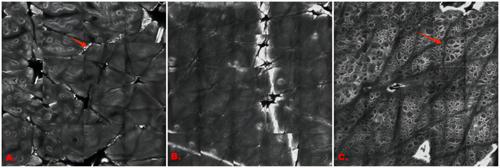
Clinical and RCM data of patients diagnosed with vitiligo were collected. RCM data were gathered from three sites of a lesion: the center of the lesion, the margin of the lesion, and the perilesional normal skin. Scoring was conducted for each method, categorizing the results as either the progressive or stable stage of vitiligo.
Results
The sensitivity was significantly lower in the perilesional normal skin than at multiple sites (p < 0.001). The specificity was significantly lower in the center of the lesion and at the margin than at multiple sites and significantly higher in the perilesional normal skin than at multiple sites (p < 0.05 for all).
Conclusions
The results of RCM of different lesion areas of vitiligo showed that the rate of missed diagnosis of the progressive stage was lowest in the center of the lesion and comparable to that of multiple sites. The misdiagnosis rate for the stable stage was lowest in the perilesional normal skin, significantly differing from the other sites. Overall, scanning multiple sites yielded the most accurate results, offering flexibility in selection according to the patient's compliance.
期刊介绍:
The Journal of Cosmetic Dermatology publishes high quality, peer-reviewed articles on all aspects of cosmetic dermatology with the aim to foster the highest standards of patient care in cosmetic dermatology. Published quarterly, the Journal of Cosmetic Dermatology facilitates continuing professional development and provides a forum for the exchange of scientific research and innovative techniques.
The scope of coverage includes, but will not be limited to: healthy skin; skin maintenance; ageing skin; photodamage and photoprotection; rejuvenation; biochemistry, endocrinology and neuroimmunology of healthy skin; imaging; skin measurement; quality of life; skin types; sensitive skin; rosacea and acne; sebum; sweat; fat; phlebology; hair conservation, restoration and removal; nails and nail surgery; pigment; psychological and medicolegal issues; retinoids; cosmetic chemistry; dermopharmacy; cosmeceuticals; toiletries; striae; cellulite; cosmetic dermatological surgery; blepharoplasty; liposuction; surgical complications; botulinum; fillers, peels and dermabrasion; local and tumescent anaesthesia; electrosurgery; lasers, including laser physics, laser research and safety, vascular lasers, pigment lasers, hair removal lasers, tattoo removal lasers, resurfacing lasers, dermal remodelling lasers and laser complications.




 京公网安备 11010802042870号
京ICP备2023020795号-1
京公网安备 11010802042870号
京ICP备2023020795号-1

 分享
分享
 求助内容:
求助内容: 应助结果提醒方式:
 扫码关注我们
