{"title":"由Mnl1/Htm1甘露糖苷酶和蛋白二硫异构酶的双功能复合体启动ERAD","authors":"Dan Zhao, Xudong Wu, Tom A. Rapoport","doi":"10.1038/s41594-025-01491-y","DOIUrl":null,"url":null,"abstract":"Misfolded glycoproteins in the endoplasmic reticulum (ER) lumen are translocated into the cytosol and degraded by the proteasome, a conserved process called ER-associated protein degradation (ERAD). In Saccharomyces cerevisiae, the glycan of these proteins is trimmed by the luminal mannosidase Mnl1 (Htm1) to generate a degradation signal. Interestingly, Mnl1 is associated with protein disulfide isomerase (Pdi1). Here we used cryo-electron microscopy, biochemical and in vivo experiments to elucidate how this complex initiates ERAD. The Mnl1–Pdi1 complex first demannosylates misfolded, globular proteins that are recognized through the C-terminal domain (CTD) of Mnl1; Pdi1 causes the CTD to ignore completely unfolded polypeptides. The disulfides of these globular proteins are then reduced by the Pdi1 component of the complex. Mnl1 blocks the canonical oxidative function of Pdi1, allowing it to function as a disulfide reductase in ERAD. The generated unfolded polypeptides can then be translocated across the membrane into the cytosol. The authors elucidate how the complex of mannosidase Mnl1 and protein disulfide isomerase (Pdi1) initiates endoplasmic-reticulum-associated protein degradation (ERAD). Mnl1 demannosylates misfolded, globular proteins and Pdi1 then reduces the disulfides to generate unfolded proteins competent for ERAD.","PeriodicalId":49141,"journal":{"name":"Nature Structural & Molecular Biology","volume":"32 6","pages":"1006-1018"},"PeriodicalIF":10.1000,"publicationDate":"2025-02-10","publicationTypes":"Journal Article","fieldsOfStudy":null,"isOpenAccess":false,"openAccessPdf":"https://www.nature.comhttps://www.nature.com/articles/s41594-025-01491-y.pdf","citationCount":"0","resultStr":"{\"title\":\"Initiation of ERAD by the bifunctional complex of Mnl1/Htm1 mannosidase and protein disulfide isomerase\",\"authors\":\"Dan Zhao, Xudong Wu, Tom A. Rapoport\",\"doi\":\"10.1038/s41594-025-01491-y\",\"DOIUrl\":null,\"url\":null,\"abstract\":\"Misfolded glycoproteins in the endoplasmic reticulum (ER) lumen are translocated into the cytosol and degraded by the proteasome, a conserved process called ER-associated protein degradation (ERAD). In Saccharomyces cerevisiae, the glycan of these proteins is trimmed by the luminal mannosidase Mnl1 (Htm1) to generate a degradation signal. Interestingly, Mnl1 is associated with protein disulfide isomerase (Pdi1). Here we used cryo-electron microscopy, biochemical and in vivo experiments to elucidate how this complex initiates ERAD. The Mnl1–Pdi1 complex first demannosylates misfolded, globular proteins that are recognized through the C-terminal domain (CTD) of Mnl1; Pdi1 causes the CTD to ignore completely unfolded polypeptides. The disulfides of these globular proteins are then reduced by the Pdi1 component of the complex. Mnl1 blocks the canonical oxidative function of Pdi1, allowing it to function as a disulfide reductase in ERAD. The generated unfolded polypeptides can then be translocated across the membrane into the cytosol. The authors elucidate how the complex of mannosidase Mnl1 and protein disulfide isomerase (Pdi1) initiates endoplasmic-reticulum-associated protein degradation (ERAD). Mnl1 demannosylates misfolded, globular proteins and Pdi1 then reduces the disulfides to generate unfolded proteins competent for ERAD.\",\"PeriodicalId\":49141,\"journal\":{\"name\":\"Nature Structural & Molecular Biology\",\"volume\":\"32 6\",\"pages\":\"1006-1018\"},\"PeriodicalIF\":10.1000,\"publicationDate\":\"2025-02-10\",\"publicationTypes\":\"Journal Article\",\"fieldsOfStudy\":null,\"isOpenAccess\":false,\"openAccessPdf\":\"https://www.nature.comhttps://www.nature.com/articles/s41594-025-01491-y.pdf\",\"citationCount\":\"0\",\"resultStr\":null,\"platform\":\"Semanticscholar\",\"paperid\":null,\"PeriodicalName\":\"Nature Structural & Molecular Biology\",\"FirstCategoryId\":\"99\",\"ListUrlMain\":\"https://www.nature.com/articles/s41594-025-01491-y\",\"RegionNum\":1,\"RegionCategory\":\"生物学\",\"ArticlePicture\":[],\"TitleCN\":null,\"AbstractTextCN\":null,\"PMCID\":null,\"EPubDate\":\"\",\"PubModel\":\"\",\"JCR\":\"Q1\",\"JCRName\":\"BIOCHEMISTRY & MOLECULAR BIOLOGY\",\"Score\":null,\"Total\":0}","platform":"Semanticscholar","paperid":null,"PeriodicalName":"Nature Structural & Molecular Biology","FirstCategoryId":"99","ListUrlMain":"https://www.nature.com/articles/s41594-025-01491-y","RegionNum":1,"RegionCategory":"生物学","ArticlePicture":[],"TitleCN":null,"AbstractTextCN":null,"PMCID":null,"EPubDate":"","PubModel":"","JCR":"Q1","JCRName":"BIOCHEMISTRY & MOLECULAR BIOLOGY","Score":null,"Total":0}
Initiation of ERAD by the bifunctional complex of Mnl1/Htm1 mannosidase and protein disulfide isomerase
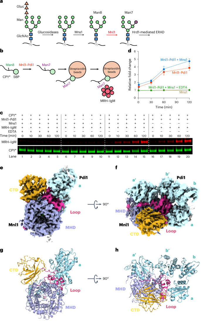
Misfolded glycoproteins in the endoplasmic reticulum (ER) lumen are translocated into the cytosol and degraded by the proteasome, a conserved process called ER-associated protein degradation (ERAD). In Saccharomyces cerevisiae, the glycan of these proteins is trimmed by the luminal mannosidase Mnl1 (Htm1) to generate a degradation signal. Interestingly, Mnl1 is associated with protein disulfide isomerase (Pdi1). Here we used cryo-electron microscopy, biochemical and in vivo experiments to elucidate how this complex initiates ERAD. The Mnl1–Pdi1 complex first demannosylates misfolded, globular proteins that are recognized through the C-terminal domain (CTD) of Mnl1; Pdi1 causes the CTD to ignore completely unfolded polypeptides. The disulfides of these globular proteins are then reduced by the Pdi1 component of the complex. Mnl1 blocks the canonical oxidative function of Pdi1, allowing it to function as a disulfide reductase in ERAD. The generated unfolded polypeptides can then be translocated across the membrane into the cytosol. The authors elucidate how the complex of mannosidase Mnl1 and protein disulfide isomerase (Pdi1) initiates endoplasmic-reticulum-associated protein degradation (ERAD). Mnl1 demannosylates misfolded, globular proteins and Pdi1 then reduces the disulfides to generate unfolded proteins competent for ERAD.
期刊介绍:
Nature Structural & Molecular Biology is a comprehensive platform that combines structural and molecular research. Our journal focuses on exploring the functional and mechanistic aspects of biological processes, emphasizing how molecular components collaborate to achieve a particular function. While structural data can shed light on these insights, our publication does not require them as a prerequisite.


 分享
分享
 求助内容:
求助内容: 应助结果提醒方式:
 扫码关注我们
