Zahra Baninameh, Jens O Watzlawik, Xu Hou, Tyrique Richardson, Nicholas W Kurchaba, Tingxiang Yan, Damian N Di Florio, DeLisa Fairweather, Lu Kang, Justin H Nguyen, Takahisa Kanekiyo, Dennis W Dickson, Sachiko Noda, Shigeto Sato, Nobutaka Hattori, Matthew S Goldberg, Ian G Ganley, Kelly L Stauch, Fabienne C Fiesel, Wolfdieter Springer
{"title":"小鼠正常衰老过程中PINK1-PRKN信号的改变。","authors":"Zahra Baninameh, Jens O Watzlawik, Xu Hou, Tyrique Richardson, Nicholas W Kurchaba, Tingxiang Yan, Damian N Di Florio, DeLisa Fairweather, Lu Kang, Justin H Nguyen, Takahisa Kanekiyo, Dennis W Dickson, Sachiko Noda, Shigeto Sato, Nobutaka Hattori, Matthew S Goldberg, Ian G Ganley, Kelly L Stauch, Fabienne C Fiesel, Wolfdieter Springer","doi":"10.1080/27694127.2024.2434379","DOIUrl":null,"url":null,"abstract":"<p><p>The ubiquitin kinase-ligase pair PINK1-PRKN identifies and selectively marks damaged mitochondria for elimination via the autophagy-lysosome system (mitophagy). While this cytoprotective pathway has been extensively studied <i>in vitro</i> upon acute and complete depolarization of mitochondria, the significance of PINK1-PRKN mitophagy <i>in vivo</i> is less well established. Here we used a novel approach to study PINK1-PRKN signaling in different energetically demanding tissues of mice during normal aging. We demonstrate a generally increased expression of both genes and enhanced enzymatic activity with aging across tissue types. Collectively our data suggest a distinct regulation of PINK1-PRKN signaling under basal conditions with the most pronounced activation and flux of the pathway in mouse heart compared to brain or skeletal muscle. Our biochemical analyses complement existing mitophagy reporter readouts and provide an important baseline assessment <i>in vivo</i>, setting the stage for further investigations of the PINK1-PRKN pathway during stress and in relevant disease conditions.</p>","PeriodicalId":72341,"journal":{"name":"Autophagy reports","volume":"3 1","pages":""},"PeriodicalIF":0.0000,"publicationDate":"2024-01-01","publicationTypes":"Journal Article","fieldsOfStudy":null,"isOpenAccess":false,"openAccessPdf":"https://www.ncbi.nlm.nih.gov/pmc/articles/PMC11855339/pdf/","citationCount":"0","resultStr":"{\"title\":\"Alterations of PINK1-PRKN signaling in mice during normal aging.\",\"authors\":\"Zahra Baninameh, Jens O Watzlawik, Xu Hou, Tyrique Richardson, Nicholas W Kurchaba, Tingxiang Yan, Damian N Di Florio, DeLisa Fairweather, Lu Kang, Justin H Nguyen, Takahisa Kanekiyo, Dennis W Dickson, Sachiko Noda, Shigeto Sato, Nobutaka Hattori, Matthew S Goldberg, Ian G Ganley, Kelly L Stauch, Fabienne C Fiesel, Wolfdieter Springer\",\"doi\":\"10.1080/27694127.2024.2434379\",\"DOIUrl\":null,\"url\":null,\"abstract\":\"<p><p>The ubiquitin kinase-ligase pair PINK1-PRKN identifies and selectively marks damaged mitochondria for elimination via the autophagy-lysosome system (mitophagy). While this cytoprotective pathway has been extensively studied <i>in vitro</i> upon acute and complete depolarization of mitochondria, the significance of PINK1-PRKN mitophagy <i>in vivo</i> is less well established. Here we used a novel approach to study PINK1-PRKN signaling in different energetically demanding tissues of mice during normal aging. We demonstrate a generally increased expression of both genes and enhanced enzymatic activity with aging across tissue types. Collectively our data suggest a distinct regulation of PINK1-PRKN signaling under basal conditions with the most pronounced activation and flux of the pathway in mouse heart compared to brain or skeletal muscle. Our biochemical analyses complement existing mitophagy reporter readouts and provide an important baseline assessment <i>in vivo</i>, setting the stage for further investigations of the PINK1-PRKN pathway during stress and in relevant disease conditions.</p>\",\"PeriodicalId\":72341,\"journal\":{\"name\":\"Autophagy reports\",\"volume\":\"3 1\",\"pages\":\"\"},\"PeriodicalIF\":0.0000,\"publicationDate\":\"2024-01-01\",\"publicationTypes\":\"Journal Article\",\"fieldsOfStudy\":null,\"isOpenAccess\":false,\"openAccessPdf\":\"https://www.ncbi.nlm.nih.gov/pmc/articles/PMC11855339/pdf/\",\"citationCount\":\"0\",\"resultStr\":null,\"platform\":\"Semanticscholar\",\"paperid\":null,\"PeriodicalName\":\"Autophagy reports\",\"FirstCategoryId\":\"1085\",\"ListUrlMain\":\"https://doi.org/10.1080/27694127.2024.2434379\",\"RegionNum\":0,\"RegionCategory\":null,\"ArticlePicture\":[],\"TitleCN\":null,\"AbstractTextCN\":null,\"PMCID\":null,\"EPubDate\":\"2024/12/7 0:00:00\",\"PubModel\":\"Epub\",\"JCR\":\"\",\"JCRName\":\"\",\"Score\":null,\"Total\":0}","platform":"Semanticscholar","paperid":null,"PeriodicalName":"Autophagy reports","FirstCategoryId":"1085","ListUrlMain":"https://doi.org/10.1080/27694127.2024.2434379","RegionNum":0,"RegionCategory":null,"ArticlePicture":[],"TitleCN":null,"AbstractTextCN":null,"PMCID":null,"EPubDate":"2024/12/7 0:00:00","PubModel":"Epub","JCR":"","JCRName":"","Score":null,"Total":0}
Alterations of PINK1-PRKN signaling in mice during normal aging.
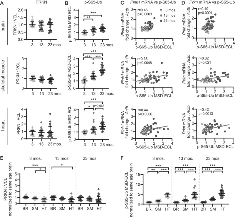
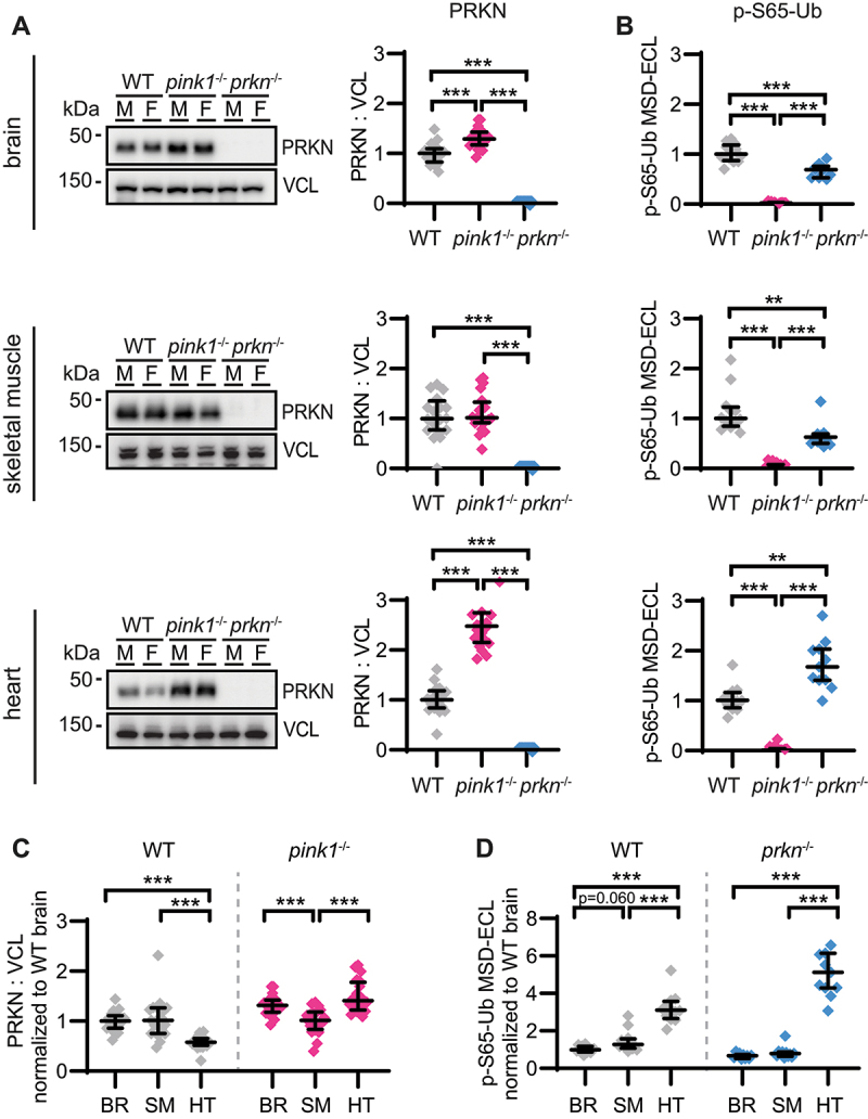
The ubiquitin kinase-ligase pair PINK1-PRKN identifies and selectively marks damaged mitochondria for elimination via the autophagy-lysosome system (mitophagy). While this cytoprotective pathway has been extensively studied in vitro upon acute and complete depolarization of mitochondria, the significance of PINK1-PRKN mitophagy in vivo is less well established. Here we used a novel approach to study PINK1-PRKN signaling in different energetically demanding tissues of mice during normal aging. We demonstrate a generally increased expression of both genes and enhanced enzymatic activity with aging across tissue types. Collectively our data suggest a distinct regulation of PINK1-PRKN signaling under basal conditions with the most pronounced activation and flux of the pathway in mouse heart compared to brain or skeletal muscle. Our biochemical analyses complement existing mitophagy reporter readouts and provide an important baseline assessment in vivo, setting the stage for further investigations of the PINK1-PRKN pathway during stress and in relevant disease conditions.


 分享
分享

 求助内容:
求助内容: 应助结果提醒方式:
 扫码关注我们
