{"title":"自然循环排列的II族内含子的结构揭示了分支和反向剪接的机制","authors":"Xiaobin Ling, Yuqi Yao, Jinbiao Ma","doi":"10.1038/s41594-025-01489-6","DOIUrl":null,"url":null,"abstract":"Circularly permuted (CP) group II introns, identified in various bacteria phyla, swap domains D5 and D6 near the 5′ end and have reversed splice sites (SSs), leading to backsplicing and circular RNA formation. In this study, we present multiple high-resolution cryo-electron microscopy structures of a natural CP group II intron from Comamonas testosteroni KF-1 (Cte 1), elucidating the molecular mechanisms of branching and backsplicing. During branching, the 5′ SS is positioned by an auxiliary sequence (AUX)-enhanced interaction between the exon-binding site and intron-binding site (IBS) and stacks on the branch-site adenosine within D6, allowing the attacking 2′-OH group to coordinate with a metal ion in the active center. In backsplicing, the 3′ SS is aligned with the branching step, leaving IBS in the active center, stabilized by base pairing with the AUX, which enables the free 3′-end hydroxyl group to directly attack the scissile phosphate of 3′ SS. Furthermore, a groove in Cte 1 may stabilize the circular RNA. These findings highlight a conserved catalytic mechanism for canonical group II introns, albeit facilitated by the versatile AUX, opening avenues for designing potent ribozymes producing circular RNAs. This study reports six cryo-electron microscopy structures of a natural group II intron, uncovering backsplicing dynamics, auxiliary sequence roles and intron space critical for circular RNA (circRNA) formation. The findings might guide improvements in circRNA production for various applications.","PeriodicalId":49141,"journal":{"name":"Nature Structural & Molecular Biology","volume":"32 6","pages":"1101-1110"},"PeriodicalIF":10.1000,"publicationDate":"2025-02-27","publicationTypes":"Journal Article","fieldsOfStudy":null,"isOpenAccess":false,"openAccessPdf":"","citationCount":"0","resultStr":"{\"title\":\"Structures of a natural circularly permuted group II intron reveal mechanisms of branching and backsplicing\",\"authors\":\"Xiaobin Ling, Yuqi Yao, Jinbiao Ma\",\"doi\":\"10.1038/s41594-025-01489-6\",\"DOIUrl\":null,\"url\":null,\"abstract\":\"Circularly permuted (CP) group II introns, identified in various bacteria phyla, swap domains D5 and D6 near the 5′ end and have reversed splice sites (SSs), leading to backsplicing and circular RNA formation. In this study, we present multiple high-resolution cryo-electron microscopy structures of a natural CP group II intron from Comamonas testosteroni KF-1 (Cte 1), elucidating the molecular mechanisms of branching and backsplicing. During branching, the 5′ SS is positioned by an auxiliary sequence (AUX)-enhanced interaction between the exon-binding site and intron-binding site (IBS) and stacks on the branch-site adenosine within D6, allowing the attacking 2′-OH group to coordinate with a metal ion in the active center. In backsplicing, the 3′ SS is aligned with the branching step, leaving IBS in the active center, stabilized by base pairing with the AUX, which enables the free 3′-end hydroxyl group to directly attack the scissile phosphate of 3′ SS. Furthermore, a groove in Cte 1 may stabilize the circular RNA. These findings highlight a conserved catalytic mechanism for canonical group II introns, albeit facilitated by the versatile AUX, opening avenues for designing potent ribozymes producing circular RNAs. This study reports six cryo-electron microscopy structures of a natural group II intron, uncovering backsplicing dynamics, auxiliary sequence roles and intron space critical for circular RNA (circRNA) formation. The findings might guide improvements in circRNA production for various applications.\",\"PeriodicalId\":49141,\"journal\":{\"name\":\"Nature Structural & Molecular Biology\",\"volume\":\"32 6\",\"pages\":\"1101-1110\"},\"PeriodicalIF\":10.1000,\"publicationDate\":\"2025-02-27\",\"publicationTypes\":\"Journal Article\",\"fieldsOfStudy\":null,\"isOpenAccess\":false,\"openAccessPdf\":\"\",\"citationCount\":\"0\",\"resultStr\":null,\"platform\":\"Semanticscholar\",\"paperid\":null,\"PeriodicalName\":\"Nature Structural & Molecular Biology\",\"FirstCategoryId\":\"99\",\"ListUrlMain\":\"https://www.nature.com/articles/s41594-025-01489-6\",\"RegionNum\":1,\"RegionCategory\":\"生物学\",\"ArticlePicture\":[],\"TitleCN\":null,\"AbstractTextCN\":null,\"PMCID\":null,\"EPubDate\":\"\",\"PubModel\":\"\",\"JCR\":\"Q1\",\"JCRName\":\"BIOCHEMISTRY & MOLECULAR BIOLOGY\",\"Score\":null,\"Total\":0}","platform":"Semanticscholar","paperid":null,"PeriodicalName":"Nature Structural & Molecular Biology","FirstCategoryId":"99","ListUrlMain":"https://www.nature.com/articles/s41594-025-01489-6","RegionNum":1,"RegionCategory":"生物学","ArticlePicture":[],"TitleCN":null,"AbstractTextCN":null,"PMCID":null,"EPubDate":"","PubModel":"","JCR":"Q1","JCRName":"BIOCHEMISTRY & MOLECULAR BIOLOGY","Score":null,"Total":0}
Structures of a natural circularly permuted group II intron reveal mechanisms of branching and backsplicing
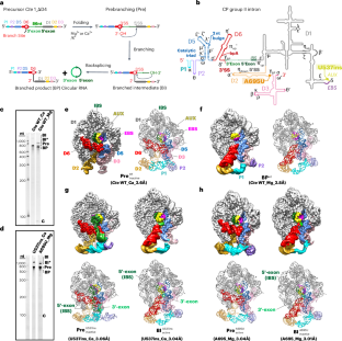
Circularly permuted (CP) group II introns, identified in various bacteria phyla, swap domains D5 and D6 near the 5′ end and have reversed splice sites (SSs), leading to backsplicing and circular RNA formation. In this study, we present multiple high-resolution cryo-electron microscopy structures of a natural CP group II intron from Comamonas testosteroni KF-1 (Cte 1), elucidating the molecular mechanisms of branching and backsplicing. During branching, the 5′ SS is positioned by an auxiliary sequence (AUX)-enhanced interaction between the exon-binding site and intron-binding site (IBS) and stacks on the branch-site adenosine within D6, allowing the attacking 2′-OH group to coordinate with a metal ion in the active center. In backsplicing, the 3′ SS is aligned with the branching step, leaving IBS in the active center, stabilized by base pairing with the AUX, which enables the free 3′-end hydroxyl group to directly attack the scissile phosphate of 3′ SS. Furthermore, a groove in Cte 1 may stabilize the circular RNA. These findings highlight a conserved catalytic mechanism for canonical group II introns, albeit facilitated by the versatile AUX, opening avenues for designing potent ribozymes producing circular RNAs. This study reports six cryo-electron microscopy structures of a natural group II intron, uncovering backsplicing dynamics, auxiliary sequence roles and intron space critical for circular RNA (circRNA) formation. The findings might guide improvements in circRNA production for various applications.
期刊介绍:
Nature Structural & Molecular Biology is a comprehensive platform that combines structural and molecular research. Our journal focuses on exploring the functional and mechanistic aspects of biological processes, emphasizing how molecular components collaborate to achieve a particular function. While structural data can shed light on these insights, our publication does not require them as a prerequisite.





 京公网安备 11010802042870号
京ICP备2023020795号-1
京公网安备 11010802042870号
京ICP备2023020795号-1

 分享
分享
 求助内容:
求助内容: 应助结果提醒方式:
 扫码关注我们
