Simone Cataldi, Paola Feraco, Maurizio Marrale, Pierpaolo Alongi, Laura Geraci, Ludovico La Grutta, Giuseppe Caruso, Tommaso Vincenzo Bartolotta, Massimo Midiri, Cesare Gagliardo
{"title":"脑胶质瘤的肿瘤内易感信号:我们站在哪里?","authors":"Simone Cataldi, Paola Feraco, Maurizio Marrale, Pierpaolo Alongi, Laura Geraci, Ludovico La Grutta, Giuseppe Caruso, Tommaso Vincenzo Bartolotta, Massimo Midiri, Cesare Gagliardo","doi":"10.3389/fradi.2025.1546069","DOIUrl":null,"url":null,"abstract":"<p><p>Nowadays, the genetic and biomolecular profile of neoplasms-related with their biological behaviour-have become a key issue in oncology, as they influence many aspects of both diagnosis and treatment. In the neuro-oncology field, neuroradiological research has recently explored the potential of non-invasively predicting the molecular phenotype of primary brain neoplasms, particularly gliomas, based on magnetic resonance imaging (MRI), using both conventional and advanced imaging techniques. Among these, diffusion-weighted imaging (DWI), perfusion-weighted imaging (PWI), MR spectroscopy (MRS) and susceptibility-weighted imaging (SWI) and have been used to explore various aspects of glioma biology, including predicting treatment response and understanding treatment-related changes during follow-up imaging. Recently, intratumoral susceptibility signals (ITSSs)-visible on SWI-have been recognised as an important new imaging tool in the evaluation of brain gliomas, as they offer a fast and simple non-invasive window into their microenvironment. These intratumoral hypointensities reflect critical pathological features such as microhemorrhages, calcifications, necrosis and vascularization. Therefore, ITSSs can provide neuroradiologists with more biological information for glioma differential diagnosis, grading and subtype differentiation, providing significant clinical support in prognosis assessment, therapeutic management and treatment response evaluation. This review summarizes recent advances in ITSS applications in glioma assessment, emphasizing both its potential and limitations while referencing key studies in the field.</p>","PeriodicalId":73101,"journal":{"name":"Frontiers in radiology","volume":"5 ","pages":"1546069"},"PeriodicalIF":2.3000,"publicationDate":"2025-02-20","publicationTypes":"Journal Article","fieldsOfStudy":null,"isOpenAccess":false,"openAccessPdf":"https://www.ncbi.nlm.nih.gov/pmc/articles/PMC11882858/pdf/","citationCount":"0","resultStr":"{\"title\":\"Intra-tumoral susceptibility signals in brain gliomas: where do we stand?\",\"authors\":\"Simone Cataldi, Paola Feraco, Maurizio Marrale, Pierpaolo Alongi, Laura Geraci, Ludovico La Grutta, Giuseppe Caruso, Tommaso Vincenzo Bartolotta, Massimo Midiri, Cesare Gagliardo\",\"doi\":\"10.3389/fradi.2025.1546069\",\"DOIUrl\":null,\"url\":null,\"abstract\":\"<p><p>Nowadays, the genetic and biomolecular profile of neoplasms-related with their biological behaviour-have become a key issue in oncology, as they influence many aspects of both diagnosis and treatment. In the neuro-oncology field, neuroradiological research has recently explored the potential of non-invasively predicting the molecular phenotype of primary brain neoplasms, particularly gliomas, based on magnetic resonance imaging (MRI), using both conventional and advanced imaging techniques. Among these, diffusion-weighted imaging (DWI), perfusion-weighted imaging (PWI), MR spectroscopy (MRS) and susceptibility-weighted imaging (SWI) and have been used to explore various aspects of glioma biology, including predicting treatment response and understanding treatment-related changes during follow-up imaging. Recently, intratumoral susceptibility signals (ITSSs)-visible on SWI-have been recognised as an important new imaging tool in the evaluation of brain gliomas, as they offer a fast and simple non-invasive window into their microenvironment. These intratumoral hypointensities reflect critical pathological features such as microhemorrhages, calcifications, necrosis and vascularization. Therefore, ITSSs can provide neuroradiologists with more biological information for glioma differential diagnosis, grading and subtype differentiation, providing significant clinical support in prognosis assessment, therapeutic management and treatment response evaluation. This review summarizes recent advances in ITSS applications in glioma assessment, emphasizing both its potential and limitations while referencing key studies in the field.</p>\",\"PeriodicalId\":73101,\"journal\":{\"name\":\"Frontiers in radiology\",\"volume\":\"5 \",\"pages\":\"1546069\"},\"PeriodicalIF\":2.3000,\"publicationDate\":\"2025-02-20\",\"publicationTypes\":\"Journal Article\",\"fieldsOfStudy\":null,\"isOpenAccess\":false,\"openAccessPdf\":\"https://www.ncbi.nlm.nih.gov/pmc/articles/PMC11882858/pdf/\",\"citationCount\":\"0\",\"resultStr\":null,\"platform\":\"Semanticscholar\",\"paperid\":null,\"PeriodicalName\":\"Frontiers in radiology\",\"FirstCategoryId\":\"1085\",\"ListUrlMain\":\"https://doi.org/10.3389/fradi.2025.1546069\",\"RegionNum\":0,\"RegionCategory\":null,\"ArticlePicture\":[],\"TitleCN\":null,\"AbstractTextCN\":null,\"PMCID\":null,\"EPubDate\":\"2025/1/1 0:00:00\",\"PubModel\":\"eCollection\",\"JCR\":\"\",\"JCRName\":\"\",\"Score\":null,\"Total\":0}","platform":"Semanticscholar","paperid":null,"PeriodicalName":"Frontiers in radiology","FirstCategoryId":"1085","ListUrlMain":"https://doi.org/10.3389/fradi.2025.1546069","RegionNum":0,"RegionCategory":null,"ArticlePicture":[],"TitleCN":null,"AbstractTextCN":null,"PMCID":null,"EPubDate":"2025/1/1 0:00:00","PubModel":"eCollection","JCR":"","JCRName":"","Score":null,"Total":0}
Intra-tumoral susceptibility signals in brain gliomas: where do we stand?
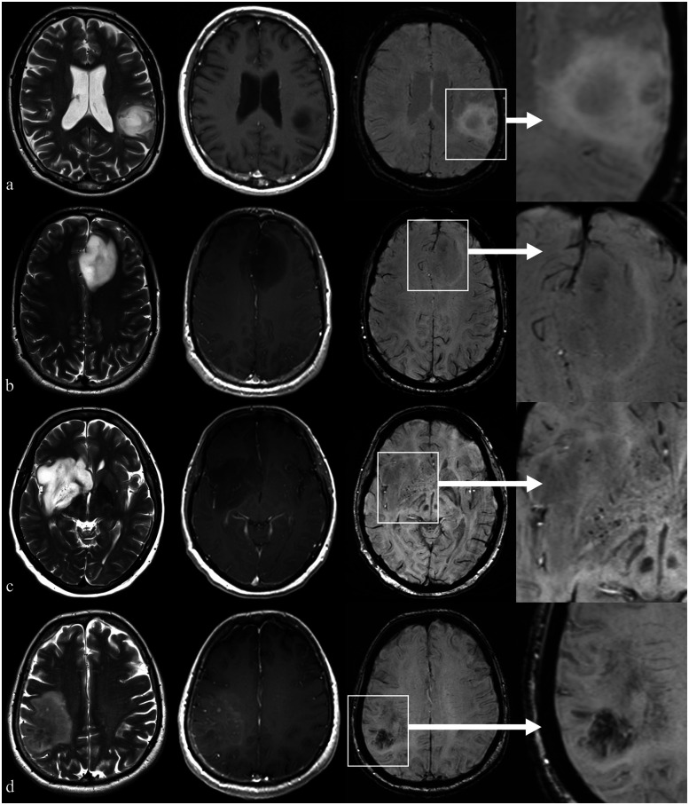
Nowadays, the genetic and biomolecular profile of neoplasms-related with their biological behaviour-have become a key issue in oncology, as they influence many aspects of both diagnosis and treatment. In the neuro-oncology field, neuroradiological research has recently explored the potential of non-invasively predicting the molecular phenotype of primary brain neoplasms, particularly gliomas, based on magnetic resonance imaging (MRI), using both conventional and advanced imaging techniques. Among these, diffusion-weighted imaging (DWI), perfusion-weighted imaging (PWI), MR spectroscopy (MRS) and susceptibility-weighted imaging (SWI) and have been used to explore various aspects of glioma biology, including predicting treatment response and understanding treatment-related changes during follow-up imaging. Recently, intratumoral susceptibility signals (ITSSs)-visible on SWI-have been recognised as an important new imaging tool in the evaluation of brain gliomas, as they offer a fast and simple non-invasive window into their microenvironment. These intratumoral hypointensities reflect critical pathological features such as microhemorrhages, calcifications, necrosis and vascularization. Therefore, ITSSs can provide neuroradiologists with more biological information for glioma differential diagnosis, grading and subtype differentiation, providing significant clinical support in prognosis assessment, therapeutic management and treatment response evaluation. This review summarizes recent advances in ITSS applications in glioma assessment, emphasizing both its potential and limitations while referencing key studies in the field.


 分享
分享

 求助内容:
求助内容: 应助结果提醒方式:
 扫码关注我们
