{"title":"量化计算机断层扫描的多个多平面重建之间的可变性。","authors":"James E Miles, Lene E Buelund","doi":"10.1186/s42490-021-00047-7","DOIUrl":null,"url":null,"abstract":"<p><strong>Background: </strong>Multiplanar reconstructions of computed tomography (CT) scans can alleviate issues with bone or joint positioning during scan acquisition. The repeatability of these reconstructions is dependent on human operators applying reconstruction criteria, and therefore is subject to error, which could affect measurement reliability for angular or spatial measurements made for orthopaedic surgery. We describe a method for quantifying inter-reconstruction variability numerically and graphically using metadata from the CT header to find vectors describing reconstruction axis alignment. The approach is demonstrated using 3 sets of computed tomographic reconstructions of 24 vulpine femorotibial joints.</p><p><strong>Results: </strong>Vectors describing axis alignments permitted identification and subsequent analysis of deviations from optimal alignment between reconstruction sets. For the worked example, alignment deviations equivalent to femoral abduction/adduction were nearly twice those for extension/flexion, and simulation of the effects of these deviations on measurements closely matched published data.</p><p><strong>Conclusions: </strong>The method presented here is straightforward and permits numerical and graphical analysis of reconstruction variability. Reconstruction alignment variability should be considered before adopting new reconstruction criteria for clinical use, and evaluated whenever there is suspicion that reconstruction variability could unduly influence subsequent measurements. These evaluations may help drive improvements in reconstruction criteria. The methods described here could also be employed for comparing patient positioning between scans and between different scan modalities.</p>","PeriodicalId":72425,"journal":{"name":"BMC biomedical engineering","volume":"3 1","pages":"2"},"PeriodicalIF":0.0000,"publicationDate":"2021-02-01","publicationTypes":"Journal Article","fieldsOfStudy":null,"isOpenAccess":false,"openAccessPdf":"https://sci-hub-pdf.com/10.1186/s42490-021-00047-7","citationCount":"0","resultStr":"{\"title\":\"Quantifying the variability between multiple multiplanar reconstructions of computed tomography scans.\",\"authors\":\"James E Miles, Lene E Buelund\",\"doi\":\"10.1186/s42490-021-00047-7\",\"DOIUrl\":null,\"url\":null,\"abstract\":\"<p><strong>Background: </strong>Multiplanar reconstructions of computed tomography (CT) scans can alleviate issues with bone or joint positioning during scan acquisition. The repeatability of these reconstructions is dependent on human operators applying reconstruction criteria, and therefore is subject to error, which could affect measurement reliability for angular or spatial measurements made for orthopaedic surgery. We describe a method for quantifying inter-reconstruction variability numerically and graphically using metadata from the CT header to find vectors describing reconstruction axis alignment. The approach is demonstrated using 3 sets of computed tomographic reconstructions of 24 vulpine femorotibial joints.</p><p><strong>Results: </strong>Vectors describing axis alignments permitted identification and subsequent analysis of deviations from optimal alignment between reconstruction sets. For the worked example, alignment deviations equivalent to femoral abduction/adduction were nearly twice those for extension/flexion, and simulation of the effects of these deviations on measurements closely matched published data.</p><p><strong>Conclusions: </strong>The method presented here is straightforward and permits numerical and graphical analysis of reconstruction variability. Reconstruction alignment variability should be considered before adopting new reconstruction criteria for clinical use, and evaluated whenever there is suspicion that reconstruction variability could unduly influence subsequent measurements. These evaluations may help drive improvements in reconstruction criteria. The methods described here could also be employed for comparing patient positioning between scans and between different scan modalities.</p>\",\"PeriodicalId\":72425,\"journal\":{\"name\":\"BMC biomedical engineering\",\"volume\":\"3 1\",\"pages\":\"2\"},\"PeriodicalIF\":0.0000,\"publicationDate\":\"2021-02-01\",\"publicationTypes\":\"Journal Article\",\"fieldsOfStudy\":null,\"isOpenAccess\":false,\"openAccessPdf\":\"https://sci-hub-pdf.com/10.1186/s42490-021-00047-7\",\"citationCount\":\"0\",\"resultStr\":null,\"platform\":\"Semanticscholar\",\"paperid\":null,\"PeriodicalName\":\"BMC biomedical engineering\",\"FirstCategoryId\":\"1085\",\"ListUrlMain\":\"https://doi.org/10.1186/s42490-021-00047-7\",\"RegionNum\":0,\"RegionCategory\":null,\"ArticlePicture\":[],\"TitleCN\":null,\"AbstractTextCN\":null,\"PMCID\":null,\"EPubDate\":\"\",\"PubModel\":\"\",\"JCR\":\"\",\"JCRName\":\"\",\"Score\":null,\"Total\":0}","platform":"Semanticscholar","paperid":null,"PeriodicalName":"BMC biomedical engineering","FirstCategoryId":"1085","ListUrlMain":"https://doi.org/10.1186/s42490-021-00047-7","RegionNum":0,"RegionCategory":null,"ArticlePicture":[],"TitleCN":null,"AbstractTextCN":null,"PMCID":null,"EPubDate":"","PubModel":"","JCR":"","JCRName":"","Score":null,"Total":0}
Quantifying the variability between multiple multiplanar reconstructions of computed tomography scans.
Background: Multiplanar reconstructions of computed tomography (CT) scans can alleviate issues with bone or joint positioning during scan acquisition. The repeatability of these reconstructions is dependent on human operators applying reconstruction criteria, and therefore is subject to error, which could affect measurement reliability for angular or spatial measurements made for orthopaedic surgery. We describe a method for quantifying inter-reconstruction variability numerically and graphically using metadata from the CT header to find vectors describing reconstruction axis alignment. The approach is demonstrated using 3 sets of computed tomographic reconstructions of 24 vulpine femorotibial joints.
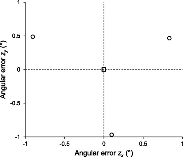
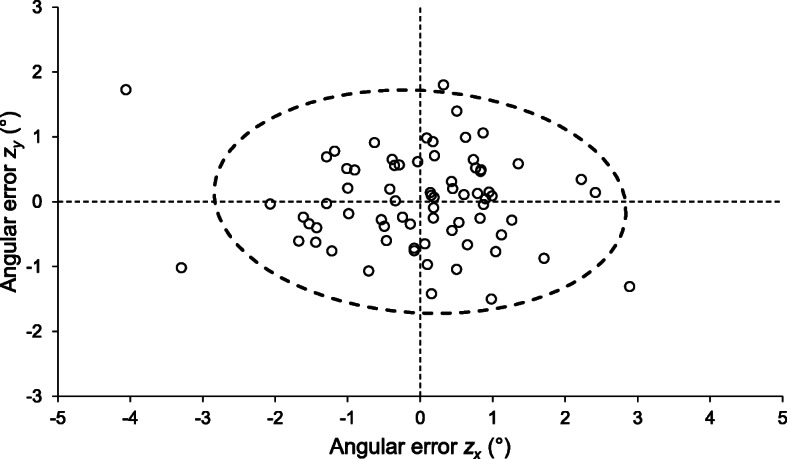
Results: Vectors describing axis alignments permitted identification and subsequent analysis of deviations from optimal alignment between reconstruction sets. For the worked example, alignment deviations equivalent to femoral abduction/adduction were nearly twice those for extension/flexion, and simulation of the effects of these deviations on measurements closely matched published data.
Conclusions: The method presented here is straightforward and permits numerical and graphical analysis of reconstruction variability. Reconstruction alignment variability should be considered before adopting new reconstruction criteria for clinical use, and evaluated whenever there is suspicion that reconstruction variability could unduly influence subsequent measurements. These evaluations may help drive improvements in reconstruction criteria. The methods described here could also be employed for comparing patient positioning between scans and between different scan modalities.


 分享
分享

 求助内容:
求助内容: 应助结果提醒方式:
 扫码关注我们
