Hidetoshi Mori, Jennifer Bolen, Louis Schuetter, Pierre Massion, Clifford C Hoyt, Scott VandenBerg, Laura Esserman, Alexander D Borowsky, Michael J Campbell
{"title":"基于酪酰胺的多重免疫荧光表征肿瘤免疫微环境。","authors":"Hidetoshi Mori, Jennifer Bolen, Louis Schuetter, Pierre Massion, Clifford C Hoyt, Scott VandenBerg, Laura Esserman, Alexander D Borowsky, Michael J Campbell","doi":"10.1007/s10911-021-09479-2","DOIUrl":null,"url":null,"abstract":"<p><p>Multiplex immunofluorescence (mIF) allows simultaneous antibody-based detection of multiple markers with a nuclear counterstain on a single tissue section. Recent studies have demonstrated that mIF is becoming an important tool for immune profiling the tumor microenvironment, further advancing our understanding of the interplay between cancer and the immune system, and identifying predictive biomarkers of response to immunotherapy. Expediting mIF discoveries is leading to improved diagnostic panels, whereas it is important that mIF protocols be standardized to facilitate their transition into clinical use. Manual processing of sections for mIF is time consuming and a potential source of variability across numerous samples. To increase reproducibility and throughput we demonstrate the use of an automated slide stainer for mIF incorporating tyramide signal amplification (TSA). We describe two panels aimed at characterizing the tumor immune microenvironment. Panel 1 included CD3, CD20, CD117, FOXP3, Ki67, pancytokeratins (CK), and DAPI, and Panel 2 included CD3, CD8, CD68, PD-1, PD-L1, CK, and DAPI. Primary antibodies were first tested by standard immunohistochemistry and single-plex IF, then multiplex panels were developed and images were obtained using a Vectra 3.0 multispectral imaging system. Various methods for image analysis (identifying cell types, determining cell densities, characterizing cell-cell associations) are outlined. These mIF protocols will be invaluable tools for immune profiling the tumor microenvironment.</p>","PeriodicalId":16413,"journal":{"name":"Journal of Mammary Gland Biology and Neoplasia","volume":"25 4","pages":"417-432"},"PeriodicalIF":3.1000,"publicationDate":"2020-12-01","publicationTypes":"Journal Article","fieldsOfStudy":null,"isOpenAccess":false,"openAccessPdf":"https://sci-hub-pdf.com/10.1007/s10911-021-09479-2","citationCount":"23","resultStr":"{\"title\":\"Characterizing the Tumor Immune Microenvironment with Tyramide-Based Multiplex Immunofluorescence.\",\"authors\":\"Hidetoshi Mori, Jennifer Bolen, Louis Schuetter, Pierre Massion, Clifford C Hoyt, Scott VandenBerg, Laura Esserman, Alexander D Borowsky, Michael J Campbell\",\"doi\":\"10.1007/s10911-021-09479-2\",\"DOIUrl\":null,\"url\":null,\"abstract\":\"<p><p>Multiplex immunofluorescence (mIF) allows simultaneous antibody-based detection of multiple markers with a nuclear counterstain on a single tissue section. Recent studies have demonstrated that mIF is becoming an important tool for immune profiling the tumor microenvironment, further advancing our understanding of the interplay between cancer and the immune system, and identifying predictive biomarkers of response to immunotherapy. Expediting mIF discoveries is leading to improved diagnostic panels, whereas it is important that mIF protocols be standardized to facilitate their transition into clinical use. Manual processing of sections for mIF is time consuming and a potential source of variability across numerous samples. To increase reproducibility and throughput we demonstrate the use of an automated slide stainer for mIF incorporating tyramide signal amplification (TSA). We describe two panels aimed at characterizing the tumor immune microenvironment. Panel 1 included CD3, CD20, CD117, FOXP3, Ki67, pancytokeratins (CK), and DAPI, and Panel 2 included CD3, CD8, CD68, PD-1, PD-L1, CK, and DAPI. Primary antibodies were first tested by standard immunohistochemistry and single-plex IF, then multiplex panels were developed and images were obtained using a Vectra 3.0 multispectral imaging system. Various methods for image analysis (identifying cell types, determining cell densities, characterizing cell-cell associations) are outlined. These mIF protocols will be invaluable tools for immune profiling the tumor microenvironment.</p>\",\"PeriodicalId\":16413,\"journal\":{\"name\":\"Journal of Mammary Gland Biology and Neoplasia\",\"volume\":\"25 4\",\"pages\":\"417-432\"},\"PeriodicalIF\":3.1000,\"publicationDate\":\"2020-12-01\",\"publicationTypes\":\"Journal Article\",\"fieldsOfStudy\":null,\"isOpenAccess\":false,\"openAccessPdf\":\"https://sci-hub-pdf.com/10.1007/s10911-021-09479-2\",\"citationCount\":\"23\",\"resultStr\":null,\"platform\":\"Semanticscholar\",\"paperid\":null,\"PeriodicalName\":\"Journal of Mammary Gland Biology and Neoplasia\",\"FirstCategoryId\":\"3\",\"ListUrlMain\":\"https://doi.org/10.1007/s10911-021-09479-2\",\"RegionNum\":4,\"RegionCategory\":\"医学\",\"ArticlePicture\":[],\"TitleCN\":null,\"AbstractTextCN\":null,\"PMCID\":null,\"EPubDate\":\"2021/2/15 0:00:00\",\"PubModel\":\"Epub\",\"JCR\":\"Q2\",\"JCRName\":\"ENDOCRINOLOGY & METABOLISM\",\"Score\":null,\"Total\":0}","platform":"Semanticscholar","paperid":null,"PeriodicalName":"Journal of Mammary Gland Biology and Neoplasia","FirstCategoryId":"3","ListUrlMain":"https://doi.org/10.1007/s10911-021-09479-2","RegionNum":4,"RegionCategory":"医学","ArticlePicture":[],"TitleCN":null,"AbstractTextCN":null,"PMCID":null,"EPubDate":"2021/2/15 0:00:00","PubModel":"Epub","JCR":"Q2","JCRName":"ENDOCRINOLOGY & METABOLISM","Score":null,"Total":0}
Characterizing the Tumor Immune Microenvironment with Tyramide-Based Multiplex Immunofluorescence.
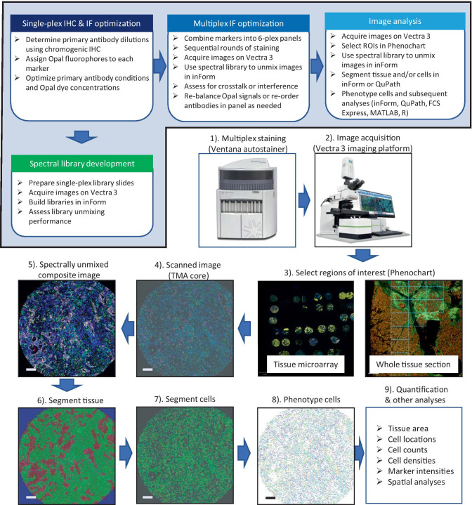
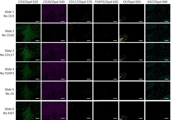
Multiplex immunofluorescence (mIF) allows simultaneous antibody-based detection of multiple markers with a nuclear counterstain on a single tissue section. Recent studies have demonstrated that mIF is becoming an important tool for immune profiling the tumor microenvironment, further advancing our understanding of the interplay between cancer and the immune system, and identifying predictive biomarkers of response to immunotherapy. Expediting mIF discoveries is leading to improved diagnostic panels, whereas it is important that mIF protocols be standardized to facilitate their transition into clinical use. Manual processing of sections for mIF is time consuming and a potential source of variability across numerous samples. To increase reproducibility and throughput we demonstrate the use of an automated slide stainer for mIF incorporating tyramide signal amplification (TSA). We describe two panels aimed at characterizing the tumor immune microenvironment. Panel 1 included CD3, CD20, CD117, FOXP3, Ki67, pancytokeratins (CK), and DAPI, and Panel 2 included CD3, CD8, CD68, PD-1, PD-L1, CK, and DAPI. Primary antibodies were first tested by standard immunohistochemistry and single-plex IF, then multiplex panels were developed and images were obtained using a Vectra 3.0 multispectral imaging system. Various methods for image analysis (identifying cell types, determining cell densities, characterizing cell-cell associations) are outlined. These mIF protocols will be invaluable tools for immune profiling the tumor microenvironment.
期刊介绍:
Journal of Mammary Gland Biology and Neoplasia is the leading Journal in the field of mammary gland biology that provides researchers within and outside the field of mammary gland biology with an integrated source of information pertaining to the development, function, and pathology of the mammary gland and its function.
Commencing in 2015, the Journal will begin receiving and publishing a combination of reviews and original, peer-reviewed research. The Journal covers all topics related to the field of mammary gland biology, including mammary development, breast cancer biology, lactation, and milk composition and quality. The environmental, endocrine, nutritional, and molecular factors regulating these processes is covered, including from a comparative biology perspective.





 京公网安备 11010802042870号
京ICP备2023020795号-1
京公网安备 11010802042870号
京ICP备2023020795号-1

 分享
分享
 求助内容:
求助内容: 应助结果提醒方式:
 扫码关注我们
