{"title":"单次静脉注射乙醇对兔肝窦内皮细胞的早期影响。","authors":"Frank Jacobs, Eddie Wisse, Bart De Geest","doi":"10.1186/1476-5926-8-4","DOIUrl":null,"url":null,"abstract":"<p><strong>Background: </strong>It has been postulated that ethanol affects hepatic sinusoidal and perisinusoidal cells. In the current experimental study, we investigated the early effect of a single intravenous dose of ethanol on the diameter of liver sinusoidal endothelial fenestrae in New Zealand White rabbits. The diameter of fenestrae in these rabbits is similar to the diameter found in humans with healthy livers. The effect of ethanol on the size of fenestrae was studied using transmission electron microscopy, because plastic embedding provides true measures for the diameter of fenestrae.</p><p><strong>Results: </strong>After intravenous administration of a single dose of 0.75 g/kg, ethanol concentration peaked at 1.1 +/- 0.10 g/l at ten minutes after injection. Compared to control rabbits (103 +/- 1.1 nm; n = 8), the average diameter of fenestrae in ethanol-injected rabbits determined at 10 minutes after injection was significantly (p < 0.01) smaller (96 +/- 2.2 nm; n = 5). Detailed analysis of distribution histograms of the diameters of fenestrae showed that the effect of ethanol was highly homogeneous.</p><p><strong>Conclusion: </strong>A decrease of the diameter of fenestrae 10 minutes after ethanol administration is likely the earliest morphological alteration induced by ethanol in the liver and underscores the potential role of liver sinusoidal endothelial cells in alcoholic liver injury.</p>","PeriodicalId":84474,"journal":{"name":"Comparative hepatology","volume":"8 ","pages":"4"},"PeriodicalIF":0.0000,"publicationDate":"2009-07-13","publicationTypes":"Journal Article","fieldsOfStudy":null,"isOpenAccess":false,"openAccessPdf":"https://sci-hub-pdf.com/10.1186/1476-5926-8-4","citationCount":"3","resultStr":"{\"title\":\"Early effect of a single intravenous injection of ethanol on hepatic sinusoidal endothelial fenestrae in rabbits.\",\"authors\":\"Frank Jacobs, Eddie Wisse, Bart De Geest\",\"doi\":\"10.1186/1476-5926-8-4\",\"DOIUrl\":null,\"url\":null,\"abstract\":\"<p><strong>Background: </strong>It has been postulated that ethanol affects hepatic sinusoidal and perisinusoidal cells. In the current experimental study, we investigated the early effect of a single intravenous dose of ethanol on the diameter of liver sinusoidal endothelial fenestrae in New Zealand White rabbits. The diameter of fenestrae in these rabbits is similar to the diameter found in humans with healthy livers. The effect of ethanol on the size of fenestrae was studied using transmission electron microscopy, because plastic embedding provides true measures for the diameter of fenestrae.</p><p><strong>Results: </strong>After intravenous administration of a single dose of 0.75 g/kg, ethanol concentration peaked at 1.1 +/- 0.10 g/l at ten minutes after injection. Compared to control rabbits (103 +/- 1.1 nm; n = 8), the average diameter of fenestrae in ethanol-injected rabbits determined at 10 minutes after injection was significantly (p < 0.01) smaller (96 +/- 2.2 nm; n = 5). Detailed analysis of distribution histograms of the diameters of fenestrae showed that the effect of ethanol was highly homogeneous.</p><p><strong>Conclusion: </strong>A decrease of the diameter of fenestrae 10 minutes after ethanol administration is likely the earliest morphological alteration induced by ethanol in the liver and underscores the potential role of liver sinusoidal endothelial cells in alcoholic liver injury.</p>\",\"PeriodicalId\":84474,\"journal\":{\"name\":\"Comparative hepatology\",\"volume\":\"8 \",\"pages\":\"4\"},\"PeriodicalIF\":0.0000,\"publicationDate\":\"2009-07-13\",\"publicationTypes\":\"Journal Article\",\"fieldsOfStudy\":null,\"isOpenAccess\":false,\"openAccessPdf\":\"https://sci-hub-pdf.com/10.1186/1476-5926-8-4\",\"citationCount\":\"3\",\"resultStr\":null,\"platform\":\"Semanticscholar\",\"paperid\":null,\"PeriodicalName\":\"Comparative hepatology\",\"FirstCategoryId\":\"1085\",\"ListUrlMain\":\"https://doi.org/10.1186/1476-5926-8-4\",\"RegionNum\":0,\"RegionCategory\":null,\"ArticlePicture\":[],\"TitleCN\":null,\"AbstractTextCN\":null,\"PMCID\":null,\"EPubDate\":\"\",\"PubModel\":\"\",\"JCR\":\"\",\"JCRName\":\"\",\"Score\":null,\"Total\":0}","platform":"Semanticscholar","paperid":null,"PeriodicalName":"Comparative hepatology","FirstCategoryId":"1085","ListUrlMain":"https://doi.org/10.1186/1476-5926-8-4","RegionNum":0,"RegionCategory":null,"ArticlePicture":[],"TitleCN":null,"AbstractTextCN":null,"PMCID":null,"EPubDate":"","PubModel":"","JCR":"","JCRName":"","Score":null,"Total":0}
Early effect of a single intravenous injection of ethanol on hepatic sinusoidal endothelial fenestrae in rabbits.
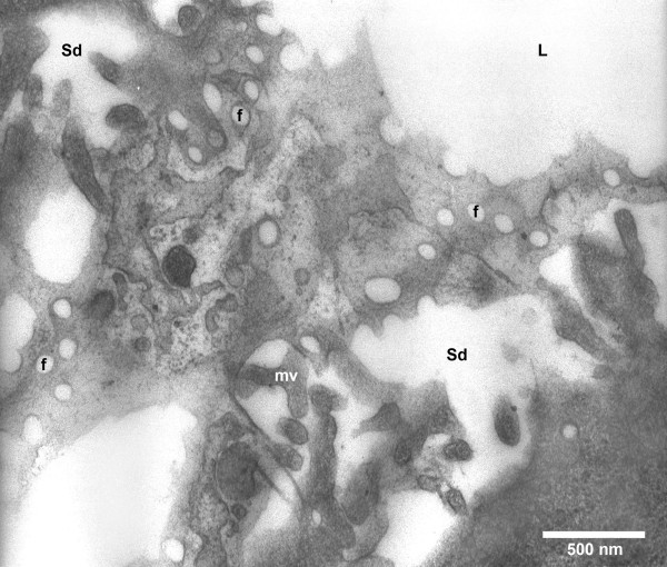
Background: It has been postulated that ethanol affects hepatic sinusoidal and perisinusoidal cells. In the current experimental study, we investigated the early effect of a single intravenous dose of ethanol on the diameter of liver sinusoidal endothelial fenestrae in New Zealand White rabbits. The diameter of fenestrae in these rabbits is similar to the diameter found in humans with healthy livers. The effect of ethanol on the size of fenestrae was studied using transmission electron microscopy, because plastic embedding provides true measures for the diameter of fenestrae.
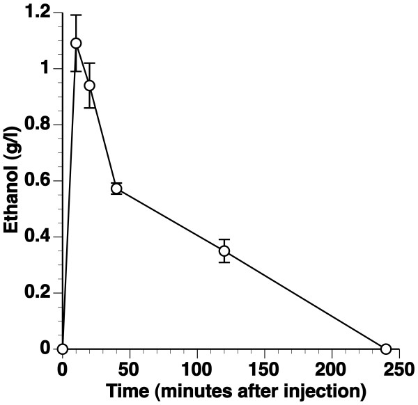
Results: After intravenous administration of a single dose of 0.75 g/kg, ethanol concentration peaked at 1.1 +/- 0.10 g/l at ten minutes after injection. Compared to control rabbits (103 +/- 1.1 nm; n = 8), the average diameter of fenestrae in ethanol-injected rabbits determined at 10 minutes after injection was significantly (p < 0.01) smaller (96 +/- 2.2 nm; n = 5). Detailed analysis of distribution histograms of the diameters of fenestrae showed that the effect of ethanol was highly homogeneous.
Conclusion: A decrease of the diameter of fenestrae 10 minutes after ethanol administration is likely the earliest morphological alteration induced by ethanol in the liver and underscores the potential role of liver sinusoidal endothelial cells in alcoholic liver injury.


 分享
分享

 求助内容:
求助内容: 应助结果提醒方式:
 扫码关注我们
