Julien Villeneuve, Fanny Pelluard-Nehme, Chantal Combe, Dominique Carles, Christine Chaponnier, Jean Ripoche, Charles Balabaud, Paulette Bioulac-Sage, Sébastien Lepreux
{"title":"正常和纤维性(管板畸形)胎儿肝门道成熟过程中间充质细胞表型变化的免疫组织化学研究。","authors":"Julien Villeneuve, Fanny Pelluard-Nehme, Chantal Combe, Dominique Carles, Christine Chaponnier, Jean Ripoche, Charles Balabaud, Paulette Bioulac-Sage, Sébastien Lepreux","doi":"10.1186/1476-5926-8-5","DOIUrl":null,"url":null,"abstract":"<p><strong>Background: </strong>In adult liver, the mesenchymal cells, portal fibroblasts and vascular smooth muscle cells can transdifferentiate into myofibroblasts, and are involved in portal fibrosis. Differential expression of markers, such as alpha-smooth muscle actin (ASMA), h-caldesmon and cellular retinol-binding protein-1 allows their phenotypic discrimination. The aim of our study was to explore the phenotypic evolution of the mesenchymal cells during fetal development in normal liver and in liver with portal fibrosis secondary to ductal plate malformation in a series of Meckel-Gruber syndrome, autosomal recessive polycystic kidney disease and Ivemark's syndrome.</p><p><strong>Results: </strong>At the early steps of the portal tract maturation, portal mesenchymal cells expressed only ASMA. During the maturation process, these cells were found condensed around the biliary and vascular structures. At the end of maturation process, only cells around vessels expressed ASMA and cells of the artery tunica media also expressed h-caldesmon. In contrast, ASMA positive cells persisted around the abnormal biliary ducts in fibrous livers.</p><p><strong>Conclusion: </strong>As in adult liver, there is a phenotypic heterogeneity of the mesenchymal cells during fetal liver development. During portal tract maturation, myofibroblastic cells disappear in normal development but persist in fibrosis following ductal plate malformation.</p>","PeriodicalId":84474,"journal":{"name":"Comparative hepatology","volume":"8 ","pages":"5"},"PeriodicalIF":0.0000,"publicationDate":"2009-07-14","publicationTypes":"Journal Article","fieldsOfStudy":null,"isOpenAccess":false,"openAccessPdf":"https://sci-hub-pdf.com/10.1186/1476-5926-8-5","citationCount":"20","resultStr":"{\"title\":\"Immunohistochemical study of the phenotypic change of the mesenchymal cells during portal tract maturation in normal and fibrous (ductal plate malformation) fetal liver.\",\"authors\":\"Julien Villeneuve, Fanny Pelluard-Nehme, Chantal Combe, Dominique Carles, Christine Chaponnier, Jean Ripoche, Charles Balabaud, Paulette Bioulac-Sage, Sébastien Lepreux\",\"doi\":\"10.1186/1476-5926-8-5\",\"DOIUrl\":null,\"url\":null,\"abstract\":\"<p><strong>Background: </strong>In adult liver, the mesenchymal cells, portal fibroblasts and vascular smooth muscle cells can transdifferentiate into myofibroblasts, and are involved in portal fibrosis. Differential expression of markers, such as alpha-smooth muscle actin (ASMA), h-caldesmon and cellular retinol-binding protein-1 allows their phenotypic discrimination. The aim of our study was to explore the phenotypic evolution of the mesenchymal cells during fetal development in normal liver and in liver with portal fibrosis secondary to ductal plate malformation in a series of Meckel-Gruber syndrome, autosomal recessive polycystic kidney disease and Ivemark's syndrome.</p><p><strong>Results: </strong>At the early steps of the portal tract maturation, portal mesenchymal cells expressed only ASMA. During the maturation process, these cells were found condensed around the biliary and vascular structures. At the end of maturation process, only cells around vessels expressed ASMA and cells of the artery tunica media also expressed h-caldesmon. In contrast, ASMA positive cells persisted around the abnormal biliary ducts in fibrous livers.</p><p><strong>Conclusion: </strong>As in adult liver, there is a phenotypic heterogeneity of the mesenchymal cells during fetal liver development. During portal tract maturation, myofibroblastic cells disappear in normal development but persist in fibrosis following ductal plate malformation.</p>\",\"PeriodicalId\":84474,\"journal\":{\"name\":\"Comparative hepatology\",\"volume\":\"8 \",\"pages\":\"5\"},\"PeriodicalIF\":0.0000,\"publicationDate\":\"2009-07-14\",\"publicationTypes\":\"Journal Article\",\"fieldsOfStudy\":null,\"isOpenAccess\":false,\"openAccessPdf\":\"https://sci-hub-pdf.com/10.1186/1476-5926-8-5\",\"citationCount\":\"20\",\"resultStr\":null,\"platform\":\"Semanticscholar\",\"paperid\":null,\"PeriodicalName\":\"Comparative hepatology\",\"FirstCategoryId\":\"1085\",\"ListUrlMain\":\"https://doi.org/10.1186/1476-5926-8-5\",\"RegionNum\":0,\"RegionCategory\":null,\"ArticlePicture\":[],\"TitleCN\":null,\"AbstractTextCN\":null,\"PMCID\":null,\"EPubDate\":\"\",\"PubModel\":\"\",\"JCR\":\"\",\"JCRName\":\"\",\"Score\":null,\"Total\":0}","platform":"Semanticscholar","paperid":null,"PeriodicalName":"Comparative hepatology","FirstCategoryId":"1085","ListUrlMain":"https://doi.org/10.1186/1476-5926-8-5","RegionNum":0,"RegionCategory":null,"ArticlePicture":[],"TitleCN":null,"AbstractTextCN":null,"PMCID":null,"EPubDate":"","PubModel":"","JCR":"","JCRName":"","Score":null,"Total":0}
Immunohistochemical study of the phenotypic change of the mesenchymal cells during portal tract maturation in normal and fibrous (ductal plate malformation) fetal liver.
Background: In adult liver, the mesenchymal cells, portal fibroblasts and vascular smooth muscle cells can transdifferentiate into myofibroblasts, and are involved in portal fibrosis. Differential expression of markers, such as alpha-smooth muscle actin (ASMA), h-caldesmon and cellular retinol-binding protein-1 allows their phenotypic discrimination. The aim of our study was to explore the phenotypic evolution of the mesenchymal cells during fetal development in normal liver and in liver with portal fibrosis secondary to ductal plate malformation in a series of Meckel-Gruber syndrome, autosomal recessive polycystic kidney disease and Ivemark's syndrome.
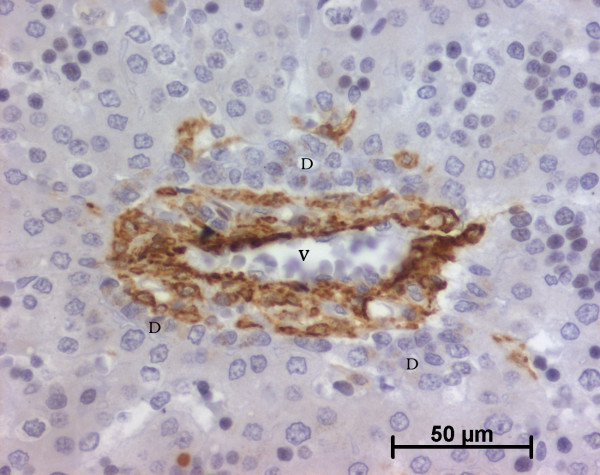
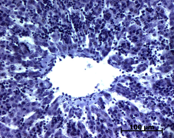
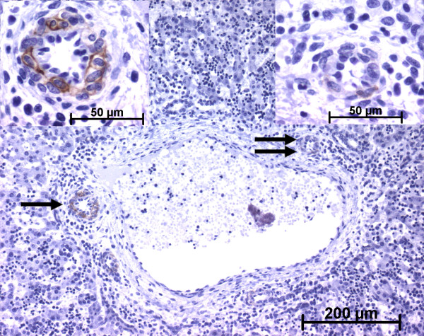
Results: At the early steps of the portal tract maturation, portal mesenchymal cells expressed only ASMA. During the maturation process, these cells were found condensed around the biliary and vascular structures. At the end of maturation process, only cells around vessels expressed ASMA and cells of the artery tunica media also expressed h-caldesmon. In contrast, ASMA positive cells persisted around the abnormal biliary ducts in fibrous livers.
Conclusion: As in adult liver, there is a phenotypic heterogeneity of the mesenchymal cells during fetal liver development. During portal tract maturation, myofibroblastic cells disappear in normal development but persist in fibrosis following ductal plate malformation.


 分享
分享

 求助内容:
求助内容: 应助结果提醒方式:
 扫码关注我们
