Yuriy K Bashmakov, Nailya A Zigangirova, Yulia P Pashko, Lidia N Kapotina, Ivan M Petyaev
{"title":"甲伐他汀对HepG2细胞沙眼衣原体生长的抑制及ldl受体水平的恢复。","authors":"Yuriy K Bashmakov, Nailya A Zigangirova, Yulia P Pashko, Lidia N Kapotina, Ivan M Petyaev","doi":"10.1186/1476-5926-9-3","DOIUrl":null,"url":null,"abstract":"<p><strong>Background: </strong>Perihepatitis is rare but consistently occurring extragenital manifestation of untreated Chlamydia trachomatis infection. Despite of possible liver involvement in generalized C. trachomatis infection, the ability of the pathogen to propagate in the hepatic cells and its impact on liver functions is not thoroughly investigated. The effect of mevastatin, an inhibitor of 3-hydroxy-3-methylglutaryl CoA reductase, on C. trachomatis growth in human hepatoma cell line HepG2 has been studied. Bacterial growth was assessed by immunostaining with FITC-labeled monoclonal antibody against chlamydial lipopolysaccharide and by RT-PCR for two chlamydial genetic markers (16S rRNA and euo).</p><p><strong>Results: </strong>Chlamydial inclusion bodies were seen in approximately 50% of hepatocytes at 48 hours in the post infection period. Lysates obtained from infected hepatocytes were positive in the infective progeny test at 48 and especially in 72 hours after infection initiation. It has been shown that chlamydial infection in hepatocytes also leads to the decline of LDL-receptor mRNA which reflects infection multiplicity rate. Additions of mevastatin (1, 20 and 40 microM) 1 hour before inoculation restored and upregulated LDL-receptor mRNA level in a dose-dependent manner. Mevastatin treatment had no effect on internalization of chlamydial particles. However it reduced drastically the number of chlamydial 16S rRNA and euo transcripts as well as overall infection rate in HepG-2 cells. Complete eradication of infection has been seen by immunofluorescent staining at 40 microM mevastatin concentration, when expression level of chlamydial 16S rRNA and euo was undetectable. Lower concentration of mevastatin (20 microM) promoted euo expression level and the appearance of atypically small chlamydial inclusions, while there was a noticeable reduction in the number of infected cells and 16S rRNA transcripts.</p><p><strong>Conclusions: </strong>C. trachomatis can efficiently propagate in hepatocytes affecting transcription rate of some liver-specific genes. Ongoing cholesterol synthesis is essential for chlamydial growth in hepatocytes. Inhibitors of cholesterol biosynthesis can supplement conventional strategy in the management of C. trachomatis infection.</p>","PeriodicalId":84474,"journal":{"name":"Comparative hepatology","volume":"9 ","pages":"3"},"PeriodicalIF":0.0000,"publicationDate":"2010-01-28","publicationTypes":"Journal Article","fieldsOfStudy":null,"isOpenAccess":false,"openAccessPdf":"https://sci-hub-pdf.com/10.1186/1476-5926-9-3","citationCount":"18","resultStr":"{\"title\":\"Chlamydia trachomatis growth inhibition and restoration of LDL-receptor level in HepG2 cells treated with mevastatin.\",\"authors\":\"Yuriy K Bashmakov, Nailya A Zigangirova, Yulia P Pashko, Lidia N Kapotina, Ivan M Petyaev\",\"doi\":\"10.1186/1476-5926-9-3\",\"DOIUrl\":null,\"url\":null,\"abstract\":\"<p><strong>Background: </strong>Perihepatitis is rare but consistently occurring extragenital manifestation of untreated Chlamydia trachomatis infection. Despite of possible liver involvement in generalized C. trachomatis infection, the ability of the pathogen to propagate in the hepatic cells and its impact on liver functions is not thoroughly investigated. The effect of mevastatin, an inhibitor of 3-hydroxy-3-methylglutaryl CoA reductase, on C. trachomatis growth in human hepatoma cell line HepG2 has been studied. Bacterial growth was assessed by immunostaining with FITC-labeled monoclonal antibody against chlamydial lipopolysaccharide and by RT-PCR for two chlamydial genetic markers (16S rRNA and euo).</p><p><strong>Results: </strong>Chlamydial inclusion bodies were seen in approximately 50% of hepatocytes at 48 hours in the post infection period. Lysates obtained from infected hepatocytes were positive in the infective progeny test at 48 and especially in 72 hours after infection initiation. It has been shown that chlamydial infection in hepatocytes also leads to the decline of LDL-receptor mRNA which reflects infection multiplicity rate. Additions of mevastatin (1, 20 and 40 microM) 1 hour before inoculation restored and upregulated LDL-receptor mRNA level in a dose-dependent manner. Mevastatin treatment had no effect on internalization of chlamydial particles. However it reduced drastically the number of chlamydial 16S rRNA and euo transcripts as well as overall infection rate in HepG-2 cells. Complete eradication of infection has been seen by immunofluorescent staining at 40 microM mevastatin concentration, when expression level of chlamydial 16S rRNA and euo was undetectable. Lower concentration of mevastatin (20 microM) promoted euo expression level and the appearance of atypically small chlamydial inclusions, while there was a noticeable reduction in the number of infected cells and 16S rRNA transcripts.</p><p><strong>Conclusions: </strong>C. trachomatis can efficiently propagate in hepatocytes affecting transcription rate of some liver-specific genes. Ongoing cholesterol synthesis is essential for chlamydial growth in hepatocytes. Inhibitors of cholesterol biosynthesis can supplement conventional strategy in the management of C. trachomatis infection.</p>\",\"PeriodicalId\":84474,\"journal\":{\"name\":\"Comparative hepatology\",\"volume\":\"9 \",\"pages\":\"3\"},\"PeriodicalIF\":0.0000,\"publicationDate\":\"2010-01-28\",\"publicationTypes\":\"Journal Article\",\"fieldsOfStudy\":null,\"isOpenAccess\":false,\"openAccessPdf\":\"https://sci-hub-pdf.com/10.1186/1476-5926-9-3\",\"citationCount\":\"18\",\"resultStr\":null,\"platform\":\"Semanticscholar\",\"paperid\":null,\"PeriodicalName\":\"Comparative hepatology\",\"FirstCategoryId\":\"1085\",\"ListUrlMain\":\"https://doi.org/10.1186/1476-5926-9-3\",\"RegionNum\":0,\"RegionCategory\":null,\"ArticlePicture\":[],\"TitleCN\":null,\"AbstractTextCN\":null,\"PMCID\":null,\"EPubDate\":\"\",\"PubModel\":\"\",\"JCR\":\"\",\"JCRName\":\"\",\"Score\":null,\"Total\":0}","platform":"Semanticscholar","paperid":null,"PeriodicalName":"Comparative hepatology","FirstCategoryId":"1085","ListUrlMain":"https://doi.org/10.1186/1476-5926-9-3","RegionNum":0,"RegionCategory":null,"ArticlePicture":[],"TitleCN":null,"AbstractTextCN":null,"PMCID":null,"EPubDate":"","PubModel":"","JCR":"","JCRName":"","Score":null,"Total":0}
Chlamydia trachomatis growth inhibition and restoration of LDL-receptor level in HepG2 cells treated with mevastatin.
Background: Perihepatitis is rare but consistently occurring extragenital manifestation of untreated Chlamydia trachomatis infection. Despite of possible liver involvement in generalized C. trachomatis infection, the ability of the pathogen to propagate in the hepatic cells and its impact on liver functions is not thoroughly investigated. The effect of mevastatin, an inhibitor of 3-hydroxy-3-methylglutaryl CoA reductase, on C. trachomatis growth in human hepatoma cell line HepG2 has been studied. Bacterial growth was assessed by immunostaining with FITC-labeled monoclonal antibody against chlamydial lipopolysaccharide and by RT-PCR for two chlamydial genetic markers (16S rRNA and euo).
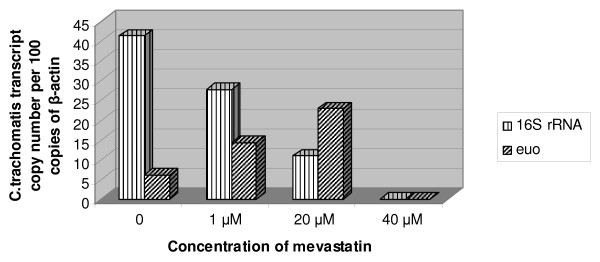
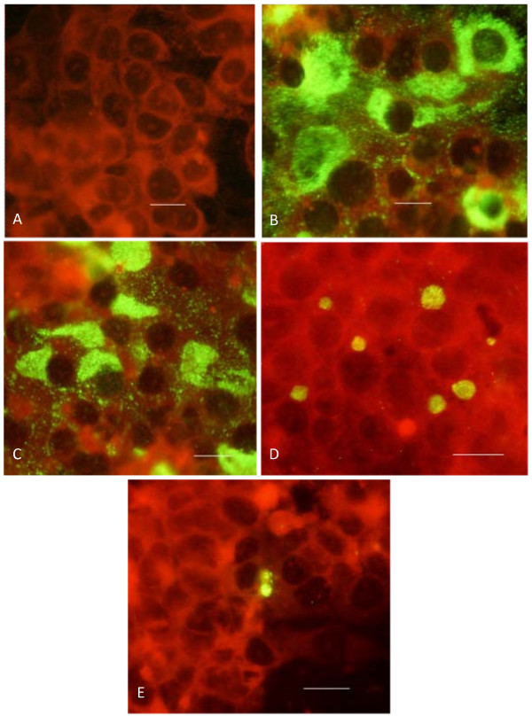
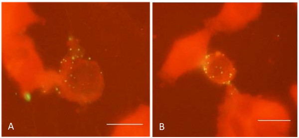
Results: Chlamydial inclusion bodies were seen in approximately 50% of hepatocytes at 48 hours in the post infection period. Lysates obtained from infected hepatocytes were positive in the infective progeny test at 48 and especially in 72 hours after infection initiation. It has been shown that chlamydial infection in hepatocytes also leads to the decline of LDL-receptor mRNA which reflects infection multiplicity rate. Additions of mevastatin (1, 20 and 40 microM) 1 hour before inoculation restored and upregulated LDL-receptor mRNA level in a dose-dependent manner. Mevastatin treatment had no effect on internalization of chlamydial particles. However it reduced drastically the number of chlamydial 16S rRNA and euo transcripts as well as overall infection rate in HepG-2 cells. Complete eradication of infection has been seen by immunofluorescent staining at 40 microM mevastatin concentration, when expression level of chlamydial 16S rRNA and euo was undetectable. Lower concentration of mevastatin (20 microM) promoted euo expression level and the appearance of atypically small chlamydial inclusions, while there was a noticeable reduction in the number of infected cells and 16S rRNA transcripts.
Conclusions: C. trachomatis can efficiently propagate in hepatocytes affecting transcription rate of some liver-specific genes. Ongoing cholesterol synthesis is essential for chlamydial growth in hepatocytes. Inhibitors of cholesterol biosynthesis can supplement conventional strategy in the management of C. trachomatis infection.


 分享
分享

 求助内容:
求助内容: 应助结果提醒方式:
 扫码关注我们
