Edmund Chinchar, Kristina L Makey, John Gibson, Fang Chen, Shelby A Cole, Gail C Megason, Srinivassan Vijayakumar, Lucio Miele, Jian-Wei Gu
{"title":"舒尼替尼显著抑制三阴性乳腺癌的增殖、迁移、抗凋亡、肿瘤血管生成和生长,但增加乳腺癌干细胞。","authors":"Edmund Chinchar, Kristina L Makey, John Gibson, Fang Chen, Shelby A Cole, Gail C Megason, Srinivassan Vijayakumar, Lucio Miele, Jian-Wei Gu","doi":"10.1186/2045-824X-6-12","DOIUrl":null,"url":null,"abstract":"<p><p>The majority of triple-negative breast cancers (TNBCs) are basal-like breast cancers. However there is no reported study on anti-tumor effects of sunitinib in xenografts of basal-like TNBC (MDA-MB-468) cells. In the present study, MDA-MB-231, MDA-MB-468, MCF-7 cells were cultured using RPMI 1640 media with 10% FBS. Vascular endothelia growth factor (VEGF) protein levels were detected using ELISA (R & D Systams). MDA-MB-468 cells were exposed to sunitinib for 18 hours for measuring proliferation (3H-thymidine incorporation), migration (BD Invasion Chamber), and apoptosis (ApopTag and ApoScreen Anuexin V Kit). The effect of sunitinib on Notch-1 expression was determined by Western blot in cultured MDA-MB-468 cells. 10(6) MDA-MB-468 cells were inoculated into the left fourth mammary gland fat pad in athymic nude-foxn1 mice. When the tumor volume reached 100 mm(3), sunitinib was given by gavage at 80 mg/kg/2 days for 4 weeks. Tumor angiogenesis was determined by CD31 immunohistochemistry. Breast cancer stem cells (CSCs) isolated from the tumors were determined by flow cytometry analysis using CD44(+)/CD24(-) or low. ELISA indicated that VEGF was much more highly expressed in MDA-MB-468 cells than MDA-MB-231 and MCF-7 cells. Sunitinib significantly inhibited the proliferation, invasion, and apoptosis resistance in cultured basal like breast cancer cells. Sunitinib significantly increased the expression of Notch-1 protein in cultured MDA-MB-468 or MDA-MB-231 cells. The xenograft models showed that oral sunitinib significantly reduced the tumor volume of TNBCs in association with the inhibition of tumor angiogeneisis, but increased breast CSCs. These findings support the hypothesis that the possibility should be considered of sunitinib increasing breast CSCs though it inhibits TNBC tumor angiogenesis and growth/progression, and that effects of sunitinib on Notch expression and hypoxia may increase breast cancer stem cells. This work provides the groundwork for an innovative therapeutic strategy in TNBC therapy by using sunitinib plus γ-secretase inhibitor to simultaneously target angiogenesis and CSC. </p>","PeriodicalId":23948,"journal":{"name":"Vascular Cell","volume":"6 ","pages":"12"},"PeriodicalIF":0.0000,"publicationDate":"2014-06-01","publicationTypes":"Journal Article","fieldsOfStudy":null,"isOpenAccess":false,"openAccessPdf":"https://sci-hub-pdf.com/10.1186/2045-824X-6-12","citationCount":"71","resultStr":"{\"title\":\"Sunitinib significantly suppresses the proliferation, migration, apoptosis resistance, tumor angiogenesis and growth of triple-negative breast cancers but increases breast cancer stem cells.\",\"authors\":\"Edmund Chinchar, Kristina L Makey, John Gibson, Fang Chen, Shelby A Cole, Gail C Megason, Srinivassan Vijayakumar, Lucio Miele, Jian-Wei Gu\",\"doi\":\"10.1186/2045-824X-6-12\",\"DOIUrl\":null,\"url\":null,\"abstract\":\"<p><p>The majority of triple-negative breast cancers (TNBCs) are basal-like breast cancers. However there is no reported study on anti-tumor effects of sunitinib in xenografts of basal-like TNBC (MDA-MB-468) cells. In the present study, MDA-MB-231, MDA-MB-468, MCF-7 cells were cultured using RPMI 1640 media with 10% FBS. Vascular endothelia growth factor (VEGF) protein levels were detected using ELISA (R & D Systams). MDA-MB-468 cells were exposed to sunitinib for 18 hours for measuring proliferation (3H-thymidine incorporation), migration (BD Invasion Chamber), and apoptosis (ApopTag and ApoScreen Anuexin V Kit). The effect of sunitinib on Notch-1 expression was determined by Western blot in cultured MDA-MB-468 cells. 10(6) MDA-MB-468 cells were inoculated into the left fourth mammary gland fat pad in athymic nude-foxn1 mice. When the tumor volume reached 100 mm(3), sunitinib was given by gavage at 80 mg/kg/2 days for 4 weeks. Tumor angiogenesis was determined by CD31 immunohistochemistry. Breast cancer stem cells (CSCs) isolated from the tumors were determined by flow cytometry analysis using CD44(+)/CD24(-) or low. ELISA indicated that VEGF was much more highly expressed in MDA-MB-468 cells than MDA-MB-231 and MCF-7 cells. Sunitinib significantly inhibited the proliferation, invasion, and apoptosis resistance in cultured basal like breast cancer cells. Sunitinib significantly increased the expression of Notch-1 protein in cultured MDA-MB-468 or MDA-MB-231 cells. The xenograft models showed that oral sunitinib significantly reduced the tumor volume of TNBCs in association with the inhibition of tumor angiogeneisis, but increased breast CSCs. These findings support the hypothesis that the possibility should be considered of sunitinib increasing breast CSCs though it inhibits TNBC tumor angiogenesis and growth/progression, and that effects of sunitinib on Notch expression and hypoxia may increase breast cancer stem cells. This work provides the groundwork for an innovative therapeutic strategy in TNBC therapy by using sunitinib plus γ-secretase inhibitor to simultaneously target angiogenesis and CSC. </p>\",\"PeriodicalId\":23948,\"journal\":{\"name\":\"Vascular Cell\",\"volume\":\"6 \",\"pages\":\"12\"},\"PeriodicalIF\":0.0000,\"publicationDate\":\"2014-06-01\",\"publicationTypes\":\"Journal Article\",\"fieldsOfStudy\":null,\"isOpenAccess\":false,\"openAccessPdf\":\"https://sci-hub-pdf.com/10.1186/2045-824X-6-12\",\"citationCount\":\"71\",\"resultStr\":null,\"platform\":\"Semanticscholar\",\"paperid\":null,\"PeriodicalName\":\"Vascular Cell\",\"FirstCategoryId\":\"1085\",\"ListUrlMain\":\"https://doi.org/10.1186/2045-824X-6-12\",\"RegionNum\":0,\"RegionCategory\":null,\"ArticlePicture\":[],\"TitleCN\":null,\"AbstractTextCN\":null,\"PMCID\":null,\"EPubDate\":\"2014/1/1 0:00:00\",\"PubModel\":\"eCollection\",\"JCR\":\"Q4\",\"JCRName\":\"Neuroscience\",\"Score\":null,\"Total\":0}","platform":"Semanticscholar","paperid":null,"PeriodicalName":"Vascular Cell","FirstCategoryId":"1085","ListUrlMain":"https://doi.org/10.1186/2045-824X-6-12","RegionNum":0,"RegionCategory":null,"ArticlePicture":[],"TitleCN":null,"AbstractTextCN":null,"PMCID":null,"EPubDate":"2014/1/1 0:00:00","PubModel":"eCollection","JCR":"Q4","JCRName":"Neuroscience","Score":null,"Total":0}
引用次数: 71
摘要
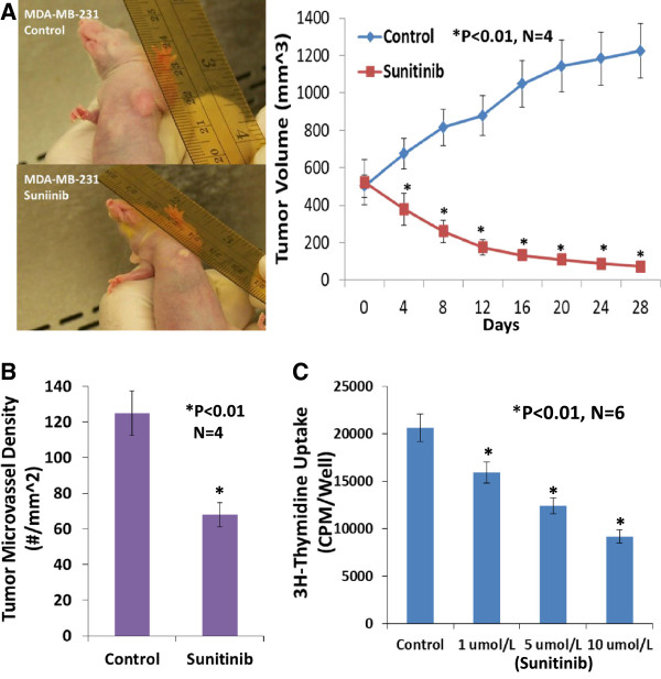
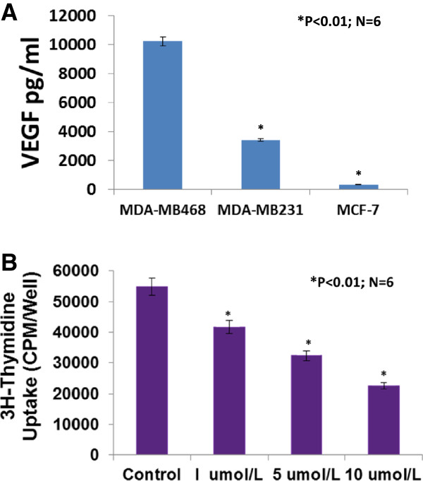
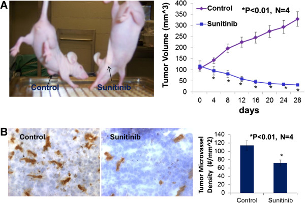
大多数三阴性乳腺癌(tnbc)是基底样乳腺癌。然而,舒尼替尼在基底样TNBC (MDA-MB-468)细胞异种移植物中的抗肿瘤作用尚未见报道。本研究采用含10%胎牛血清的RPMI 1640培养基培养MDA-MB-231、MDA-MB-468、MCF-7细胞。采用ELISA (R & D systems)检测血管内皮生长因子(VEGF)蛋白水平。将MDA-MB-468细胞暴露于舒尼替尼18小时,检测细胞增殖(3h -胸苷结合)、迁移(BD侵袭室)和凋亡(ApopTag和ApoScreen Anuexin V Kit)。Western blot检测舒尼替尼对MDA-MB-468细胞Notch-1表达的影响。10(6)将MDA-MB-468细胞接种于胸腺裸foxn1小鼠左第四乳腺脂肪垫。当肿瘤体积达到100 mm(3)时,以80 mg/kg/2天的剂量灌胃给予舒尼替尼,连续4周。CD31免疫组化检测肿瘤血管生成。用CD44(+)/CD24(-)或低水平流式细胞术检测从肿瘤中分离的乳腺癌干细胞(CSCs)。ELISA结果显示,VEGF在MDA-MB-468细胞中的表达明显高于MDA-MB-231和MCF-7细胞。舒尼替尼显著抑制培养基底样乳腺癌细胞的增殖、侵袭和抗凋亡能力。舒尼替尼显著提高MDA-MB-468或MDA-MB-231细胞中Notch-1蛋白的表达。异种移植模型显示,口服舒尼替尼显著减少tnbc的肿瘤体积,抑制肿瘤血管生成,但增加乳腺CSCs。这些发现支持了这样的假设,即应该考虑舒尼替尼通过抑制TNBC肿瘤血管生成和生长/进展来增加乳腺CSCs的可能性,舒尼替尼对Notch表达和缺氧的影响可能增加乳腺癌干细胞。本研究为利用舒尼替尼联合γ-分泌酶抑制剂同时靶向血管生成和CSC的TNBC治疗创新治疗策略奠定了基础。
Sunitinib significantly suppresses the proliferation, migration, apoptosis resistance, tumor angiogenesis and growth of triple-negative breast cancers but increases breast cancer stem cells.
The majority of triple-negative breast cancers (TNBCs) are basal-like breast cancers. However there is no reported study on anti-tumor effects of sunitinib in xenografts of basal-like TNBC (MDA-MB-468) cells. In the present study, MDA-MB-231, MDA-MB-468, MCF-7 cells were cultured using RPMI 1640 media with 10% FBS. Vascular endothelia growth factor (VEGF) protein levels were detected using ELISA (R & D Systams). MDA-MB-468 cells were exposed to sunitinib for 18 hours for measuring proliferation (3H-thymidine incorporation), migration (BD Invasion Chamber), and apoptosis (ApopTag and ApoScreen Anuexin V Kit). The effect of sunitinib on Notch-1 expression was determined by Western blot in cultured MDA-MB-468 cells. 10(6) MDA-MB-468 cells were inoculated into the left fourth mammary gland fat pad in athymic nude-foxn1 mice. When the tumor volume reached 100 mm(3), sunitinib was given by gavage at 80 mg/kg/2 days for 4 weeks. Tumor angiogenesis was determined by CD31 immunohistochemistry. Breast cancer stem cells (CSCs) isolated from the tumors were determined by flow cytometry analysis using CD44(+)/CD24(-) or low. ELISA indicated that VEGF was much more highly expressed in MDA-MB-468 cells than MDA-MB-231 and MCF-7 cells. Sunitinib significantly inhibited the proliferation, invasion, and apoptosis resistance in cultured basal like breast cancer cells. Sunitinib significantly increased the expression of Notch-1 protein in cultured MDA-MB-468 or MDA-MB-231 cells. The xenograft models showed that oral sunitinib significantly reduced the tumor volume of TNBCs in association with the inhibition of tumor angiogeneisis, but increased breast CSCs. These findings support the hypothesis that the possibility should be considered of sunitinib increasing breast CSCs though it inhibits TNBC tumor angiogenesis and growth/progression, and that effects of sunitinib on Notch expression and hypoxia may increase breast cancer stem cells. This work provides the groundwork for an innovative therapeutic strategy in TNBC therapy by using sunitinib plus γ-secretase inhibitor to simultaneously target angiogenesis and CSC.


 分享
分享
 求助内容:
求助内容: 应助结果提醒方式:
 扫码关注我们
