{"title":"接受左甲状腺素治疗的绝经前甲状腺功能减退症女性是否需要进行骨状况监测?","authors":"Ruby P Babu, Alap Christy, Anupama Hegde, Poornima Manjrekar, Vivian D'Souza","doi":"10.4137/CMWH.S22114","DOIUrl":null,"url":null,"abstract":"<p><strong>Background: </strong>Suppressive doses of levothyroxine therapy are reported to reduce bone mineral density (BMD) in women. Data on bone changes in premenopausal hypothyroid women with replacement therapy are limited. Hence, this study was undertaken to evaluate bone changes in this group using bone markers and BMD.</p><p><strong>Materials and methods: </strong>A hospital-based case-control study including 75 premenopausal women aged 30-45 years was conducted. The subjects were categorized based on their thyroid function and history into three groups of 25 euthyroid, 25 newly diagnosed hypothyroid, and 25 hypothyroid women on 100-200 μg of levothyroxine for a minimum of 5 years. The bone changes were evaluated and compared among the groups biochemically by estimating their plasma osteocalcin and serum calcium and phosphorus and radiologically by measuring their BMD by quantitative ultrasonography. Statistical analysis was conducted by using analysis of variance, Tukey's test, and Pearson's correlation using IBM SPSS Statistics 20.</p><p><strong>Results: </strong>Levels of plasma osteocalcin, serum calcium, and serum phosphorus in patients on long-term levothyroxine therapy were significantly higher than those in newly diagnosed hypothyroid women and in the euthyroid group. BMD showed definite features of osteopenia (T-score: -2.26 ± 0.5) among the women in the treatment group, while it was well within the normal range in the newly diagnosed and euthyroid women. A significant correlation was found between the osteocalcin levels and T-score.</p><p><strong>Conclusion: </strong>Hypothyroid women on long-term levothyroxine therapy showed signs of increased bone turnover and increased resorptive changes, though not frank osteoporosis. Hence, it may be important to evaluate the bone status of patients on levothyroxine for >5 years.</p>","PeriodicalId":90142,"journal":{"name":"Clinical medicine insights. Women's health","volume":"8 ","pages":"1-6"},"PeriodicalIF":0.0000,"publicationDate":"2015-03-15","publicationTypes":"Journal Article","fieldsOfStudy":null,"isOpenAccess":false,"openAccessPdf":"https://www.ncbi.nlm.nih.gov/pmc/articles/PMC4362625/pdf/","citationCount":"0","resultStr":"{\"title\":\"Do premenopausal hypothyroid women on levothyroxine therapy need bone status monitoring?\",\"authors\":\"Ruby P Babu, Alap Christy, Anupama Hegde, Poornima Manjrekar, Vivian D'Souza\",\"doi\":\"10.4137/CMWH.S22114\",\"DOIUrl\":null,\"url\":null,\"abstract\":\"<p><strong>Background: </strong>Suppressive doses of levothyroxine therapy are reported to reduce bone mineral density (BMD) in women. Data on bone changes in premenopausal hypothyroid women with replacement therapy are limited. Hence, this study was undertaken to evaluate bone changes in this group using bone markers and BMD.</p><p><strong>Materials and methods: </strong>A hospital-based case-control study including 75 premenopausal women aged 30-45 years was conducted. The subjects were categorized based on their thyroid function and history into three groups of 25 euthyroid, 25 newly diagnosed hypothyroid, and 25 hypothyroid women on 100-200 μg of levothyroxine for a minimum of 5 years. The bone changes were evaluated and compared among the groups biochemically by estimating their plasma osteocalcin and serum calcium and phosphorus and radiologically by measuring their BMD by quantitative ultrasonography. Statistical analysis was conducted by using analysis of variance, Tukey's test, and Pearson's correlation using IBM SPSS Statistics 20.</p><p><strong>Results: </strong>Levels of plasma osteocalcin, serum calcium, and serum phosphorus in patients on long-term levothyroxine therapy were significantly higher than those in newly diagnosed hypothyroid women and in the euthyroid group. BMD showed definite features of osteopenia (T-score: -2.26 ± 0.5) among the women in the treatment group, while it was well within the normal range in the newly diagnosed and euthyroid women. A significant correlation was found between the osteocalcin levels and T-score.</p><p><strong>Conclusion: </strong>Hypothyroid women on long-term levothyroxine therapy showed signs of increased bone turnover and increased resorptive changes, though not frank osteoporosis. Hence, it may be important to evaluate the bone status of patients on levothyroxine for >5 years.</p>\",\"PeriodicalId\":90142,\"journal\":{\"name\":\"Clinical medicine insights. Women's health\",\"volume\":\"8 \",\"pages\":\"1-6\"},\"PeriodicalIF\":0.0000,\"publicationDate\":\"2015-03-15\",\"publicationTypes\":\"Journal Article\",\"fieldsOfStudy\":null,\"isOpenAccess\":false,\"openAccessPdf\":\"https://www.ncbi.nlm.nih.gov/pmc/articles/PMC4362625/pdf/\",\"citationCount\":\"0\",\"resultStr\":null,\"platform\":\"Semanticscholar\",\"paperid\":null,\"PeriodicalName\":\"Clinical medicine insights. Women's health\",\"FirstCategoryId\":\"1085\",\"ListUrlMain\":\"https://doi.org/10.4137/CMWH.S22114\",\"RegionNum\":0,\"RegionCategory\":null,\"ArticlePicture\":[],\"TitleCN\":null,\"AbstractTextCN\":null,\"PMCID\":null,\"EPubDate\":\"2015/1/1 0:00:00\",\"PubModel\":\"eCollection\",\"JCR\":\"\",\"JCRName\":\"\",\"Score\":null,\"Total\":0}","platform":"Semanticscholar","paperid":null,"PeriodicalName":"Clinical medicine insights. Women's health","FirstCategoryId":"1085","ListUrlMain":"https://doi.org/10.4137/CMWH.S22114","RegionNum":0,"RegionCategory":null,"ArticlePicture":[],"TitleCN":null,"AbstractTextCN":null,"PMCID":null,"EPubDate":"2015/1/1 0:00:00","PubModel":"eCollection","JCR":"","JCRName":"","Score":null,"Total":0}
Do premenopausal hypothyroid women on levothyroxine therapy need bone status monitoring?
Background: Suppressive doses of levothyroxine therapy are reported to reduce bone mineral density (BMD) in women. Data on bone changes in premenopausal hypothyroid women with replacement therapy are limited. Hence, this study was undertaken to evaluate bone changes in this group using bone markers and BMD.
Materials and methods: A hospital-based case-control study including 75 premenopausal women aged 30-45 years was conducted. The subjects were categorized based on their thyroid function and history into three groups of 25 euthyroid, 25 newly diagnosed hypothyroid, and 25 hypothyroid women on 100-200 μg of levothyroxine for a minimum of 5 years. The bone changes were evaluated and compared among the groups biochemically by estimating their plasma osteocalcin and serum calcium and phosphorus and radiologically by measuring their BMD by quantitative ultrasonography. Statistical analysis was conducted by using analysis of variance, Tukey's test, and Pearson's correlation using IBM SPSS Statistics 20.
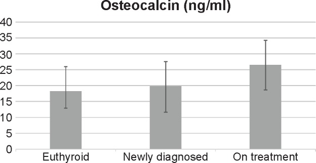
Results: Levels of plasma osteocalcin, serum calcium, and serum phosphorus in patients on long-term levothyroxine therapy were significantly higher than those in newly diagnosed hypothyroid women and in the euthyroid group. BMD showed definite features of osteopenia (T-score: -2.26 ± 0.5) among the women in the treatment group, while it was well within the normal range in the newly diagnosed and euthyroid women. A significant correlation was found between the osteocalcin levels and T-score.
Conclusion: Hypothyroid women on long-term levothyroxine therapy showed signs of increased bone turnover and increased resorptive changes, though not frank osteoporosis. Hence, it may be important to evaluate the bone status of patients on levothyroxine for >5 years.


 分享
分享

 求助内容:
求助内容: 应助结果提醒方式:
 扫码关注我们
