Luz M Morán, Jesús Vega, Nieves Gómez-León, Ana Royuela
{"title":"四肢黏液瘤和黏液样脂肪肉瘤:我们在常规、灌注和扩散磁共振中的初步发现。","authors":"Luz M Morán, Jesús Vega, Nieves Gómez-León, Ana Royuela","doi":"10.1177/20584601221131481","DOIUrl":null,"url":null,"abstract":"<p><strong>Background: </strong>The differentiation between myxomas and myxoid liposarcomas (MLPS) often is a serious challenge for the radiologists. Magnetic resonance imaging (MRI) is the most useful imaging technique in characterization of the soft tissue tumors (STT).</p><p><strong>Purpose: </strong>To evaluate in a sample of myxomas and MLPS of the extremities, what morphological findings in conventional MRI allow us to differentiate these two types of myxoid tumors, in addition to analyzing the validity of the apparent diffusion coefficient (ADC) values of diffusion-weighted MRI (DW-MRI).</p><p><strong>Material and methods: </strong>Magnetic resonance imaging studies in myxomas and MLPS of extremities searched in our PACS between 2015 and 2019. All studies had conventional MRI with T1, T2, and PD SPAIR sequences, while DW-MRI with ADC mapping and perfusion MRI with a T1 sequence repeated for 4 minutes after contrast injection were additional sequences only in some explorations. Two radiologists evaluated independently the MRI studies by examining the qualitative parameters. Apparent diffusion coefficient values were calculated using two methods-ADC global and ADC solid, and Receiver Operating Characteristic (ROC) curves were applied for analysis.</p><p><strong>Results: </strong>The features were consistent with MLPS: size greater than 10 cm, heterogeneous signal on T1, and nodular enhancement, while the common findings for myxomas were a homogenously hypointense signal on T1 and diffuse peritumoral enhancement. The solid and global ADC values were higher in myxomas. We observed that the solid ADC value less than 2.06 x 10<sup>-3</sup>mm<sup>2</sup> x s would support the diagnosis of MLPS against myxoma.</p><p><strong>Conclusion: </strong>Overall, MRI with its different modalities improved the diagnostic accuracy when differentiating myxomas from MLPS of extremities.</p>","PeriodicalId":72063,"journal":{"name":"Acta radiologica open","volume":"11 10","pages":"20584601221131481"},"PeriodicalIF":1.0000,"publicationDate":"2022-10-07","publicationTypes":"Journal Article","fieldsOfStudy":null,"isOpenAccess":false,"openAccessPdf":"https://ftp.ncbi.nlm.nih.gov/pub/pmc/oa_pdf/a9/a7/10.1177_20584601221131481.PMC9549112.pdf","citationCount":"2","resultStr":"{\"title\":\"Myxomas and myxoid liposarcomas of the extremities: Our preliminary findings in conventional, perfusion, and diffusion magnetic resonance.\",\"authors\":\"Luz M Morán, Jesús Vega, Nieves Gómez-León, Ana Royuela\",\"doi\":\"10.1177/20584601221131481\",\"DOIUrl\":null,\"url\":null,\"abstract\":\"<p><strong>Background: </strong>The differentiation between myxomas and myxoid liposarcomas (MLPS) often is a serious challenge for the radiologists. Magnetic resonance imaging (MRI) is the most useful imaging technique in characterization of the soft tissue tumors (STT).</p><p><strong>Purpose: </strong>To evaluate in a sample of myxomas and MLPS of the extremities, what morphological findings in conventional MRI allow us to differentiate these two types of myxoid tumors, in addition to analyzing the validity of the apparent diffusion coefficient (ADC) values of diffusion-weighted MRI (DW-MRI).</p><p><strong>Material and methods: </strong>Magnetic resonance imaging studies in myxomas and MLPS of extremities searched in our PACS between 2015 and 2019. All studies had conventional MRI with T1, T2, and PD SPAIR sequences, while DW-MRI with ADC mapping and perfusion MRI with a T1 sequence repeated for 4 minutes after contrast injection were additional sequences only in some explorations. Two radiologists evaluated independently the MRI studies by examining the qualitative parameters. Apparent diffusion coefficient values were calculated using two methods-ADC global and ADC solid, and Receiver Operating Characteristic (ROC) curves were applied for analysis.</p><p><strong>Results: </strong>The features were consistent with MLPS: size greater than 10 cm, heterogeneous signal on T1, and nodular enhancement, while the common findings for myxomas were a homogenously hypointense signal on T1 and diffuse peritumoral enhancement. The solid and global ADC values were higher in myxomas. We observed that the solid ADC value less than 2.06 x 10<sup>-3</sup>mm<sup>2</sup> x s would support the diagnosis of MLPS against myxoma.</p><p><strong>Conclusion: </strong>Overall, MRI with its different modalities improved the diagnostic accuracy when differentiating myxomas from MLPS of extremities.</p>\",\"PeriodicalId\":72063,\"journal\":{\"name\":\"Acta radiologica open\",\"volume\":\"11 10\",\"pages\":\"20584601221131481\"},\"PeriodicalIF\":1.0000,\"publicationDate\":\"2022-10-07\",\"publicationTypes\":\"Journal Article\",\"fieldsOfStudy\":null,\"isOpenAccess\":false,\"openAccessPdf\":\"https://ftp.ncbi.nlm.nih.gov/pub/pmc/oa_pdf/a9/a7/10.1177_20584601221131481.PMC9549112.pdf\",\"citationCount\":\"2\",\"resultStr\":null,\"platform\":\"Semanticscholar\",\"paperid\":null,\"PeriodicalName\":\"Acta radiologica open\",\"FirstCategoryId\":\"1085\",\"ListUrlMain\":\"https://doi.org/10.1177/20584601221131481\",\"RegionNum\":0,\"RegionCategory\":null,\"ArticlePicture\":[],\"TitleCN\":null,\"AbstractTextCN\":null,\"PMCID\":null,\"EPubDate\":\"2022/10/1 0:00:00\",\"PubModel\":\"eCollection\",\"JCR\":\"Q4\",\"JCRName\":\"RADIOLOGY, NUCLEAR MEDICINE & MEDICAL IMAGING\",\"Score\":null,\"Total\":0}","platform":"Semanticscholar","paperid":null,"PeriodicalName":"Acta radiologica open","FirstCategoryId":"1085","ListUrlMain":"https://doi.org/10.1177/20584601221131481","RegionNum":0,"RegionCategory":null,"ArticlePicture":[],"TitleCN":null,"AbstractTextCN":null,"PMCID":null,"EPubDate":"2022/10/1 0:00:00","PubModel":"eCollection","JCR":"Q4","JCRName":"RADIOLOGY, NUCLEAR MEDICINE & MEDICAL IMAGING","Score":null,"Total":0}
Myxomas and myxoid liposarcomas of the extremities: Our preliminary findings in conventional, perfusion, and diffusion magnetic resonance.
Background: The differentiation between myxomas and myxoid liposarcomas (MLPS) often is a serious challenge for the radiologists. Magnetic resonance imaging (MRI) is the most useful imaging technique in characterization of the soft tissue tumors (STT).
Purpose: To evaluate in a sample of myxomas and MLPS of the extremities, what morphological findings in conventional MRI allow us to differentiate these two types of myxoid tumors, in addition to analyzing the validity of the apparent diffusion coefficient (ADC) values of diffusion-weighted MRI (DW-MRI).
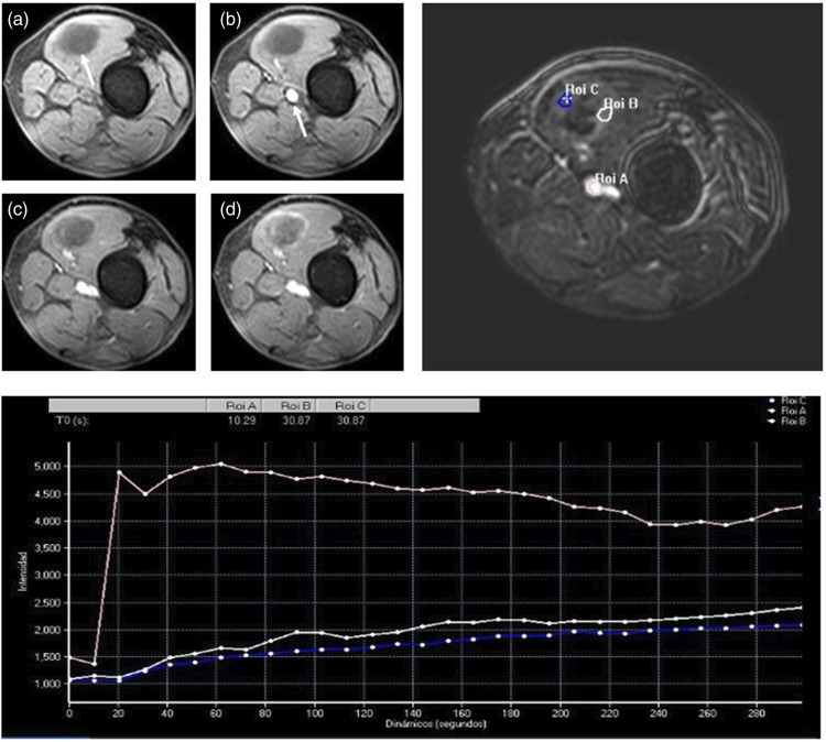
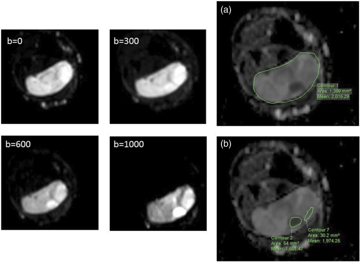
Material and methods: Magnetic resonance imaging studies in myxomas and MLPS of extremities searched in our PACS between 2015 and 2019. All studies had conventional MRI with T1, T2, and PD SPAIR sequences, while DW-MRI with ADC mapping and perfusion MRI with a T1 sequence repeated for 4 minutes after contrast injection were additional sequences only in some explorations. Two radiologists evaluated independently the MRI studies by examining the qualitative parameters. Apparent diffusion coefficient values were calculated using two methods-ADC global and ADC solid, and Receiver Operating Characteristic (ROC) curves were applied for analysis.
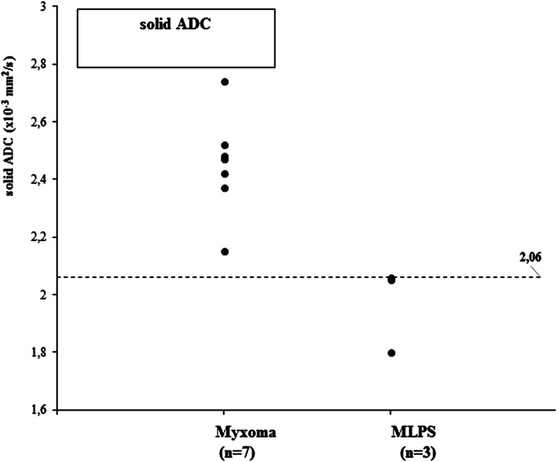
Results: The features were consistent with MLPS: size greater than 10 cm, heterogeneous signal on T1, and nodular enhancement, while the common findings for myxomas were a homogenously hypointense signal on T1 and diffuse peritumoral enhancement. The solid and global ADC values were higher in myxomas. We observed that the solid ADC value less than 2.06 x 10⁻³mm2 x s would support the diagnosis of MLPS against myxoma.
Conclusion: Overall, MRI with its different modalities improved the diagnostic accuracy when differentiating myxomas from MLPS of extremities.


 分享
分享

 求助内容:
求助内容: 应助结果提醒方式:
 扫码关注我们
