Dong Hee Ryu, Ro Hyun Sung, Min Ho Kang, Jae Woon Choi
{"title":"胰腺淋巴上皮囊肿似恶性囊性肿瘤1例报告。","authors":"Dong Hee Ryu, Ro Hyun Sung, Min Ho Kang, Jae Woon Choi","doi":"10.14701/kjhbps.2015.19.3.129","DOIUrl":null,"url":null,"abstract":"<p><p>Lymphoepithelial cysts of the pancreas are a type of true cyst that can mimic pseudocysts and cystic neoplasms. They are very rare, non-malignant lesions that are unilocular or multilocular cystic lesions lined predominantly by mature squamous epithelium and surrounded by non-neoplastic lymphoid elements. We, herein, present a patient with a cystic pancreas tumor mimicking a malignant cystic neoplasm. The patient was admitted with upper abdominal discomfort. Computed tomography showed a 64×39 mm cystic mass in the pancreas tail. She underwent distal pancreatectomy and splenectomy. In the fluid analysis of the pancreas cystic mass, the CEA and CA19-9 were 618 ng/ml and 3.9 U/ml, respectively. The resected pancreas specimen showed a 6.5 cm-sized cyst the pancreas tail. The cyst was well circumscribed and multilocular. The final pathology report of the resected pancreas specimen noted that the cyst was multilocular, and the cyst lining was showing stratified squamous epithelium covering the lymphoid tissue (containing lymphoid follicles), which was consistent with a lymphoepithelial cyst. The patient recovered uneventfully from surgery and has been doing well for the past 3 months. A differential diagnosis of cystic pancreatic lesions is important. We suggest that lymphoepithelial cysts, although very rare, may be included in the differential diagnosis of cystic pancreatic tumors. </p>","PeriodicalId":91136,"journal":{"name":"Korean journal of hepato-biliary-pancreatic surgery","volume":"19 3","pages":"129-32"},"PeriodicalIF":0.0000,"publicationDate":"2015-08-01","publicationTypes":"Journal Article","fieldsOfStudy":null,"isOpenAccess":false,"openAccessPdf":"https://sci-hub-pdf.com/10.14701/kjhbps.2015.19.3.129","citationCount":"6","resultStr":"{\"title\":\"Lymphoepithelial cyst of the pancreas mimicking malignant cystic tumor: report of a case.\",\"authors\":\"Dong Hee Ryu, Ro Hyun Sung, Min Ho Kang, Jae Woon Choi\",\"doi\":\"10.14701/kjhbps.2015.19.3.129\",\"DOIUrl\":null,\"url\":null,\"abstract\":\"<p><p>Lymphoepithelial cysts of the pancreas are a type of true cyst that can mimic pseudocysts and cystic neoplasms. They are very rare, non-malignant lesions that are unilocular or multilocular cystic lesions lined predominantly by mature squamous epithelium and surrounded by non-neoplastic lymphoid elements. We, herein, present a patient with a cystic pancreas tumor mimicking a malignant cystic neoplasm. The patient was admitted with upper abdominal discomfort. Computed tomography showed a 64×39 mm cystic mass in the pancreas tail. She underwent distal pancreatectomy and splenectomy. In the fluid analysis of the pancreas cystic mass, the CEA and CA19-9 were 618 ng/ml and 3.9 U/ml, respectively. The resected pancreas specimen showed a 6.5 cm-sized cyst the pancreas tail. The cyst was well circumscribed and multilocular. The final pathology report of the resected pancreas specimen noted that the cyst was multilocular, and the cyst lining was showing stratified squamous epithelium covering the lymphoid tissue (containing lymphoid follicles), which was consistent with a lymphoepithelial cyst. The patient recovered uneventfully from surgery and has been doing well for the past 3 months. A differential diagnosis of cystic pancreatic lesions is important. We suggest that lymphoepithelial cysts, although very rare, may be included in the differential diagnosis of cystic pancreatic tumors. </p>\",\"PeriodicalId\":91136,\"journal\":{\"name\":\"Korean journal of hepato-biliary-pancreatic surgery\",\"volume\":\"19 3\",\"pages\":\"129-32\"},\"PeriodicalIF\":0.0000,\"publicationDate\":\"2015-08-01\",\"publicationTypes\":\"Journal Article\",\"fieldsOfStudy\":null,\"isOpenAccess\":false,\"openAccessPdf\":\"https://sci-hub-pdf.com/10.14701/kjhbps.2015.19.3.129\",\"citationCount\":\"6\",\"resultStr\":null,\"platform\":\"Semanticscholar\",\"paperid\":null,\"PeriodicalName\":\"Korean journal of hepato-biliary-pancreatic surgery\",\"FirstCategoryId\":\"1085\",\"ListUrlMain\":\"https://doi.org/10.14701/kjhbps.2015.19.3.129\",\"RegionNum\":0,\"RegionCategory\":null,\"ArticlePicture\":[],\"TitleCN\":null,\"AbstractTextCN\":null,\"PMCID\":null,\"EPubDate\":\"2015/8/28 0:00:00\",\"PubModel\":\"Epub\",\"JCR\":\"\",\"JCRName\":\"\",\"Score\":null,\"Total\":0}","platform":"Semanticscholar","paperid":null,"PeriodicalName":"Korean journal of hepato-biliary-pancreatic surgery","FirstCategoryId":"1085","ListUrlMain":"https://doi.org/10.14701/kjhbps.2015.19.3.129","RegionNum":0,"RegionCategory":null,"ArticlePicture":[],"TitleCN":null,"AbstractTextCN":null,"PMCID":null,"EPubDate":"2015/8/28 0:00:00","PubModel":"Epub","JCR":"","JCRName":"","Score":null,"Total":0}
Lymphoepithelial cyst of the pancreas mimicking malignant cystic tumor: report of a case.
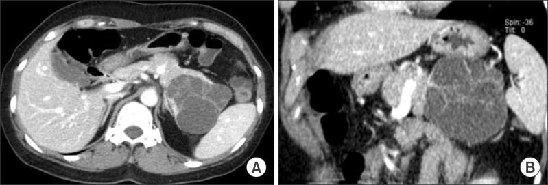
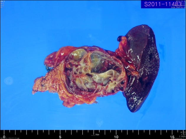
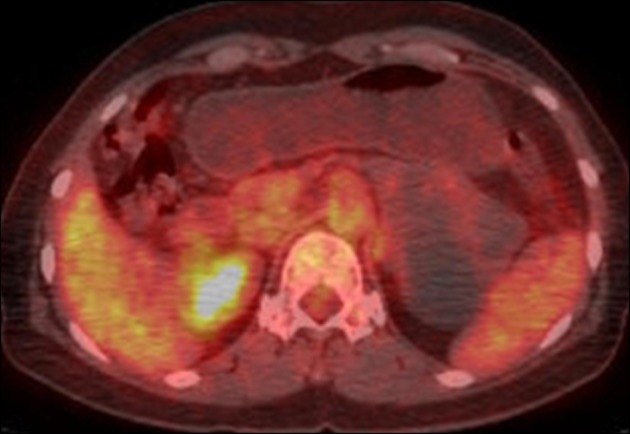
Lymphoepithelial cysts of the pancreas are a type of true cyst that can mimic pseudocysts and cystic neoplasms. They are very rare, non-malignant lesions that are unilocular or multilocular cystic lesions lined predominantly by mature squamous epithelium and surrounded by non-neoplastic lymphoid elements. We, herein, present a patient with a cystic pancreas tumor mimicking a malignant cystic neoplasm. The patient was admitted with upper abdominal discomfort. Computed tomography showed a 64×39 mm cystic mass in the pancreas tail. She underwent distal pancreatectomy and splenectomy. In the fluid analysis of the pancreas cystic mass, the CEA and CA19-9 were 618 ng/ml and 3.9 U/ml, respectively. The resected pancreas specimen showed a 6.5 cm-sized cyst the pancreas tail. The cyst was well circumscribed and multilocular. The final pathology report of the resected pancreas specimen noted that the cyst was multilocular, and the cyst lining was showing stratified squamous epithelium covering the lymphoid tissue (containing lymphoid follicles), which was consistent with a lymphoepithelial cyst. The patient recovered uneventfully from surgery and has been doing well for the past 3 months. A differential diagnosis of cystic pancreatic lesions is important. We suggest that lymphoepithelial cysts, although very rare, may be included in the differential diagnosis of cystic pancreatic tumors.


 分享
分享

 求助内容:
求助内容: 应助结果提醒方式:
 扫码关注我们
