Jason Pui Yin Cheung, Karen Ka Man Ng, Prudence Wing Hang Cheung, Dino Samartzis, Kenneth Man Chee Cheung
{"title":"腰椎发育性椎管狭窄的影像学指标。","authors":"Jason Pui Yin Cheung, Karen Ka Man Ng, Prudence Wing Hang Cheung, Dino Samartzis, Kenneth Man Chee Cheung","doi":"10.1186/s13013-017-0113-3","DOIUrl":null,"url":null,"abstract":"<p><strong>Background: </strong>Patients with developmental spinal stenosis (DSS) are susceptible to developing symptomatic stenosis due to pre-existing narrowed spinal canals. DSS has been previously defined by MRI via the axial anteroposterior (AP) bony spinal canal diameter. However, MRI is hardly a cost-efficient tool for screening patients. X-rays are superior due to its availability and cost, but currently, there is no definition of DSS based on plain radiographs. Thus, the aim of this study is to develop radiographic indices for diagnosing DSS.</p><p><strong>Methods: </strong>This was a prospective cohort of 148 subjects consisting of patients undergoing surgery for lumbar spinal stenosis (patient group) and asymptomatic subjects recruited openly from the general population (control group). Ethics approval was obtained from the local institutional review board. All subjects underwent MRI for diagnosing DSS and radiographs for measuring parameters used for creating the indices. All measurements were performed by two independent investigators, blinded to patient details. Intra- and interobserver reliability analyses were conducted, and only parameters with near perfect intraclass correlation underwent receiver operating characteristic (ROC) analysis to determine the cutoff values for diagnosing DSS using radiographs.</p><p><strong>Results: </strong>Imaging parameters from a total of 66 subjects from the patient group and 82 asymptomatic subjects in the control group were used for analysis. ROC analysis suggested sagittal vertebral body width to pedicle width ratio (SBW:PW) as having the strongest sensitivity and specificity for diagnosing DSS. Cutoff indices for SBW:PW were level-specific: L1 (2.0), L2 (2.0), L3 (2.2), L4 (2.2), L5 (2.5), and S1 (2.8).</p><p><strong>Conclusions: </strong>This is the first study to define DSS on plain radiographs based on comparisons between a clinically relevant patient group and a control group. Individuals with DSS can be identified by a simple radiograph using a screening tool allowing for better cost-saving means for clinical diagnosis or research purposes.</p>","PeriodicalId":21573,"journal":{"name":"Scoliosis and Spinal Disorders","volume":"12 ","pages":"3"},"PeriodicalIF":0.0000,"publicationDate":"2017-02-20","publicationTypes":"Journal Article","fieldsOfStudy":null,"isOpenAccess":false,"openAccessPdf":"https://sci-hub-pdf.com/10.1186/s13013-017-0113-3","citationCount":"16","resultStr":"{\"title\":\"Radiographic indices for lumbar developmental spinal stenosis.\",\"authors\":\"Jason Pui Yin Cheung, Karen Ka Man Ng, Prudence Wing Hang Cheung, Dino Samartzis, Kenneth Man Chee Cheung\",\"doi\":\"10.1186/s13013-017-0113-3\",\"DOIUrl\":null,\"url\":null,\"abstract\":\"<p><strong>Background: </strong>Patients with developmental spinal stenosis (DSS) are susceptible to developing symptomatic stenosis due to pre-existing narrowed spinal canals. DSS has been previously defined by MRI via the axial anteroposterior (AP) bony spinal canal diameter. However, MRI is hardly a cost-efficient tool for screening patients. X-rays are superior due to its availability and cost, but currently, there is no definition of DSS based on plain radiographs. Thus, the aim of this study is to develop radiographic indices for diagnosing DSS.</p><p><strong>Methods: </strong>This was a prospective cohort of 148 subjects consisting of patients undergoing surgery for lumbar spinal stenosis (patient group) and asymptomatic subjects recruited openly from the general population (control group). Ethics approval was obtained from the local institutional review board. All subjects underwent MRI for diagnosing DSS and radiographs for measuring parameters used for creating the indices. All measurements were performed by two independent investigators, blinded to patient details. Intra- and interobserver reliability analyses were conducted, and only parameters with near perfect intraclass correlation underwent receiver operating characteristic (ROC) analysis to determine the cutoff values for diagnosing DSS using radiographs.</p><p><strong>Results: </strong>Imaging parameters from a total of 66 subjects from the patient group and 82 asymptomatic subjects in the control group were used for analysis. ROC analysis suggested sagittal vertebral body width to pedicle width ratio (SBW:PW) as having the strongest sensitivity and specificity for diagnosing DSS. Cutoff indices for SBW:PW were level-specific: L1 (2.0), L2 (2.0), L3 (2.2), L4 (2.2), L5 (2.5), and S1 (2.8).</p><p><strong>Conclusions: </strong>This is the first study to define DSS on plain radiographs based on comparisons between a clinically relevant patient group and a control group. Individuals with DSS can be identified by a simple radiograph using a screening tool allowing for better cost-saving means for clinical diagnosis or research purposes.</p>\",\"PeriodicalId\":21573,\"journal\":{\"name\":\"Scoliosis and Spinal Disorders\",\"volume\":\"12 \",\"pages\":\"3\"},\"PeriodicalIF\":0.0000,\"publicationDate\":\"2017-02-20\",\"publicationTypes\":\"Journal Article\",\"fieldsOfStudy\":null,\"isOpenAccess\":false,\"openAccessPdf\":\"https://sci-hub-pdf.com/10.1186/s13013-017-0113-3\",\"citationCount\":\"16\",\"resultStr\":null,\"platform\":\"Semanticscholar\",\"paperid\":null,\"PeriodicalName\":\"Scoliosis and Spinal Disorders\",\"FirstCategoryId\":\"1085\",\"ListUrlMain\":\"https://doi.org/10.1186/s13013-017-0113-3\",\"RegionNum\":0,\"RegionCategory\":null,\"ArticlePicture\":[],\"TitleCN\":null,\"AbstractTextCN\":null,\"PMCID\":null,\"EPubDate\":\"2017/1/1 0:00:00\",\"PubModel\":\"eCollection\",\"JCR\":\"Q1\",\"JCRName\":\"Medicine\",\"Score\":null,\"Total\":0}","platform":"Semanticscholar","paperid":null,"PeriodicalName":"Scoliosis and Spinal Disorders","FirstCategoryId":"1085","ListUrlMain":"https://doi.org/10.1186/s13013-017-0113-3","RegionNum":0,"RegionCategory":null,"ArticlePicture":[],"TitleCN":null,"AbstractTextCN":null,"PMCID":null,"EPubDate":"2017/1/1 0:00:00","PubModel":"eCollection","JCR":"Q1","JCRName":"Medicine","Score":null,"Total":0}
Radiographic indices for lumbar developmental spinal stenosis.
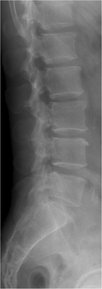
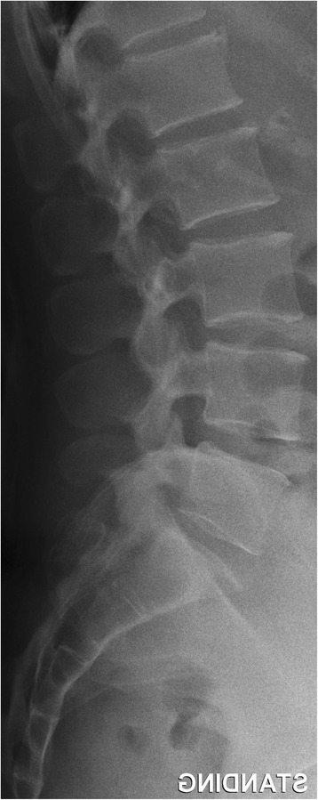
Background: Patients with developmental spinal stenosis (DSS) are susceptible to developing symptomatic stenosis due to pre-existing narrowed spinal canals. DSS has been previously defined by MRI via the axial anteroposterior (AP) bony spinal canal diameter. However, MRI is hardly a cost-efficient tool for screening patients. X-rays are superior due to its availability and cost, but currently, there is no definition of DSS based on plain radiographs. Thus, the aim of this study is to develop radiographic indices for diagnosing DSS.
Methods: This was a prospective cohort of 148 subjects consisting of patients undergoing surgery for lumbar spinal stenosis (patient group) and asymptomatic subjects recruited openly from the general population (control group). Ethics approval was obtained from the local institutional review board. All subjects underwent MRI for diagnosing DSS and radiographs for measuring parameters used for creating the indices. All measurements were performed by two independent investigators, blinded to patient details. Intra- and interobserver reliability analyses were conducted, and only parameters with near perfect intraclass correlation underwent receiver operating characteristic (ROC) analysis to determine the cutoff values for diagnosing DSS using radiographs.
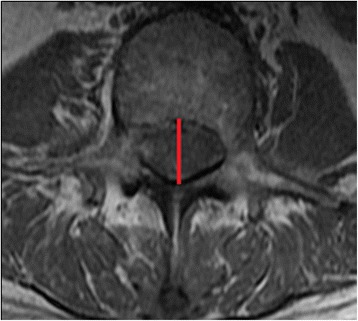
Results: Imaging parameters from a total of 66 subjects from the patient group and 82 asymptomatic subjects in the control group were used for analysis. ROC analysis suggested sagittal vertebral body width to pedicle width ratio (SBW:PW) as having the strongest sensitivity and specificity for diagnosing DSS. Cutoff indices for SBW:PW were level-specific: L1 (2.0), L2 (2.0), L3 (2.2), L4 (2.2), L5 (2.5), and S1 (2.8).
Conclusions: This is the first study to define DSS on plain radiographs based on comparisons between a clinically relevant patient group and a control group. Individuals with DSS can be identified by a simple radiograph using a screening tool allowing for better cost-saving means for clinical diagnosis or research purposes.
期刊介绍:
Cessation.Scoliosis and Spinal Disorders is an open access, multidisciplinary journal that encompasses all aspects of research on prevention, diagnosis, treatment, outcomes and cost-analyses of conservative and surgical management of all spinal deformities and disorders. Both clinical and basic science reports form the cornerstone of the journal in its endeavour to provide original, primary studies as well as narrative/systematic reviews and meta-analyses to the academic community and beyond. Scoliosis and Spinal Disorders aims to provide an integrated and balanced view of cutting-edge spine research to further enhance effective collaboration among clinical spine specialists and scientists, and to ultimately improve patient outcomes based on an evidence-based spine care approach.


 分享
分享
 求助内容:
求助内容: 应助结果提醒方式:
 扫码关注我们
