Rob C Brink, Dino Colo, Tom P C Schlösser, Koen L Vincken, Marijn van Stralen, Steve C N Hui, Lin Shi, Winnie C W Chu, Jack C Y Cheng, René M Castelein
{"title":"青少年特发性脊柱侧凸的直立、俯卧和仰卧脊柱形态和排列。","authors":"Rob C Brink, Dino Colo, Tom P C Schlösser, Koen L Vincken, Marijn van Stralen, Steve C N Hui, Lin Shi, Winnie C W Chu, Jack C Y Cheng, René M Castelein","doi":"10.1186/s13013-017-0111-5","DOIUrl":null,"url":null,"abstract":"<p><strong>Background: </strong>Patients with adolescent idiopathic scoliosis (AIS) are usually investigated by serial imaging studies during the course of treatment, some imaging involves ionizing radiation, and the radiation doses are cumulative. Few studies have addressed the correlation of spinal deformity captured by these different imaging modalities, for which patient positioning are different. To the best of our knowledge, this is the first study to compare the coronal, axial, and sagittal morphology of the scoliotic spine in three different body positions (upright, prone, and supine) and between three different imaging modalities (X-ray, CT, and MRI).</p><p><strong>Methods: </strong>Sixty-two AIS patients scheduled for scoliosis surgery, and having undergone standard pre-operative work-up, were included. This work-up included upright full-spine radiographs, supine bending radiographs, supine MRI, and prone CT as is the routine in one of our institutions. In all three positions, Cobb angles, thoracic kyphosis (TK), lumbar lordosis (LL), and vertebral rotation were determined. The relationship among three positions (upright X-ray, prone CT, and supine MRI) was investigated according to the Bland-Altman test, whereas the correlation was described by the intraclass correlation coefficient (ICC).</p><p><strong>Results: </strong>Thoracic and lumbar Cobb angles correlated significantly between conventional radiographs (68° ± 15° and 44° ± 17°), prone CT (54° ± 15° and 33° ± 15°), and supine MRI (57° ± 14° and 35° ± 16°; ICC ≥0.96; <i>P</i> < 0.001). The thoracic and lumbar apical vertebral rotation showed a good correlation among three positions (upright, 22° ± 12° and 11° ± 13°; prone, 20° ± 9° and 8° ± 11°; supine, 16° ± 11° and 6° ± 14°; ICC ≥0.82; <i>P</i> < 0.001). The TK and LL correlated well among three different positions (TK 26° ± 11°, 22° ± 12°, and 17° ± 10°; <i>P</i> ≤ 0.004; LL 49° ± 12°, 45° ± 11°, and 44° ± 12°; <i>P <</i> 0.006; ICC 0.87 and 0.85).</p><p><strong>Conclusions: </strong>Although there is a generalized underestimation of morphological parameters of the scoliotic deformity in the supine and prone positions as compared to the upright position, a significant correlation of these parameters is still evident among different body positions by different imaging modalities. Findings of this study suggest that severity of scoliotic deformity in AIS patients can be largely represented by different imaging modalities despite the difference in body positioning.</p>","PeriodicalId":21573,"journal":{"name":"Scoliosis and Spinal Disorders","volume":"12 ","pages":"6"},"PeriodicalIF":0.0000,"publicationDate":"2017-02-22","publicationTypes":"Journal Article","fieldsOfStudy":null,"isOpenAccess":false,"openAccessPdf":"https://www.ncbi.nlm.nih.gov/pmc/articles/PMC5320720/pdf/","citationCount":"0","resultStr":"{\"title\":\"Upright, prone, and supine spinal morphology and alignment in adolescent idiopathic scoliosis.\",\"authors\":\"Rob C Brink, Dino Colo, Tom P C Schlösser, Koen L Vincken, Marijn van Stralen, Steve C N Hui, Lin Shi, Winnie C W Chu, Jack C Y Cheng, René M Castelein\",\"doi\":\"10.1186/s13013-017-0111-5\",\"DOIUrl\":null,\"url\":null,\"abstract\":\"<p><strong>Background: </strong>Patients with adolescent idiopathic scoliosis (AIS) are usually investigated by serial imaging studies during the course of treatment, some imaging involves ionizing radiation, and the radiation doses are cumulative. Few studies have addressed the correlation of spinal deformity captured by these different imaging modalities, for which patient positioning are different. To the best of our knowledge, this is the first study to compare the coronal, axial, and sagittal morphology of the scoliotic spine in three different body positions (upright, prone, and supine) and between three different imaging modalities (X-ray, CT, and MRI).</p><p><strong>Methods: </strong>Sixty-two AIS patients scheduled for scoliosis surgery, and having undergone standard pre-operative work-up, were included. This work-up included upright full-spine radiographs, supine bending radiographs, supine MRI, and prone CT as is the routine in one of our institutions. In all three positions, Cobb angles, thoracic kyphosis (TK), lumbar lordosis (LL), and vertebral rotation were determined. The relationship among three positions (upright X-ray, prone CT, and supine MRI) was investigated according to the Bland-Altman test, whereas the correlation was described by the intraclass correlation coefficient (ICC).</p><p><strong>Results: </strong>Thoracic and lumbar Cobb angles correlated significantly between conventional radiographs (68° ± 15° and 44° ± 17°), prone CT (54° ± 15° and 33° ± 15°), and supine MRI (57° ± 14° and 35° ± 16°; ICC ≥0.96; <i>P</i> < 0.001). The thoracic and lumbar apical vertebral rotation showed a good correlation among three positions (upright, 22° ± 12° and 11° ± 13°; prone, 20° ± 9° and 8° ± 11°; supine, 16° ± 11° and 6° ± 14°; ICC ≥0.82; <i>P</i> < 0.001). The TK and LL correlated well among three different positions (TK 26° ± 11°, 22° ± 12°, and 17° ± 10°; <i>P</i> ≤ 0.004; LL 49° ± 12°, 45° ± 11°, and 44° ± 12°; <i>P <</i> 0.006; ICC 0.87 and 0.85).</p><p><strong>Conclusions: </strong>Although there is a generalized underestimation of morphological parameters of the scoliotic deformity in the supine and prone positions as compared to the upright position, a significant correlation of these parameters is still evident among different body positions by different imaging modalities. Findings of this study suggest that severity of scoliotic deformity in AIS patients can be largely represented by different imaging modalities despite the difference in body positioning.</p>\",\"PeriodicalId\":21573,\"journal\":{\"name\":\"Scoliosis and Spinal Disorders\",\"volume\":\"12 \",\"pages\":\"6\"},\"PeriodicalIF\":0.0000,\"publicationDate\":\"2017-02-22\",\"publicationTypes\":\"Journal Article\",\"fieldsOfStudy\":null,\"isOpenAccess\":false,\"openAccessPdf\":\"https://www.ncbi.nlm.nih.gov/pmc/articles/PMC5320720/pdf/\",\"citationCount\":\"0\",\"resultStr\":null,\"platform\":\"Semanticscholar\",\"paperid\":null,\"PeriodicalName\":\"Scoliosis and Spinal Disorders\",\"FirstCategoryId\":\"1085\",\"ListUrlMain\":\"https://doi.org/10.1186/s13013-017-0111-5\",\"RegionNum\":0,\"RegionCategory\":null,\"ArticlePicture\":[],\"TitleCN\":null,\"AbstractTextCN\":null,\"PMCID\":null,\"EPubDate\":\"2017/1/1 0:00:00\",\"PubModel\":\"eCollection\",\"JCR\":\"Q1\",\"JCRName\":\"Medicine\",\"Score\":null,\"Total\":0}","platform":"Semanticscholar","paperid":null,"PeriodicalName":"Scoliosis and Spinal Disorders","FirstCategoryId":"1085","ListUrlMain":"https://doi.org/10.1186/s13013-017-0111-5","RegionNum":0,"RegionCategory":null,"ArticlePicture":[],"TitleCN":null,"AbstractTextCN":null,"PMCID":null,"EPubDate":"2017/1/1 0:00:00","PubModel":"eCollection","JCR":"Q1","JCRName":"Medicine","Score":null,"Total":0}
Upright, prone, and supine spinal morphology and alignment in adolescent idiopathic scoliosis.
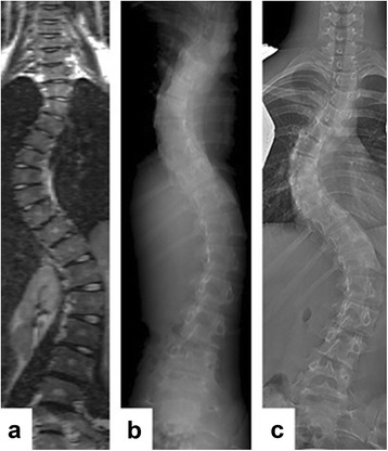
Background: Patients with adolescent idiopathic scoliosis (AIS) are usually investigated by serial imaging studies during the course of treatment, some imaging involves ionizing radiation, and the radiation doses are cumulative. Few studies have addressed the correlation of spinal deformity captured by these different imaging modalities, for which patient positioning are different. To the best of our knowledge, this is the first study to compare the coronal, axial, and sagittal morphology of the scoliotic spine in three different body positions (upright, prone, and supine) and between three different imaging modalities (X-ray, CT, and MRI).
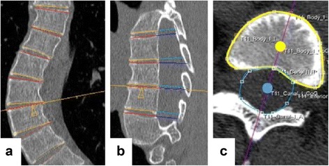
Methods: Sixty-two AIS patients scheduled for scoliosis surgery, and having undergone standard pre-operative work-up, were included. This work-up included upright full-spine radiographs, supine bending radiographs, supine MRI, and prone CT as is the routine in one of our institutions. In all three positions, Cobb angles, thoracic kyphosis (TK), lumbar lordosis (LL), and vertebral rotation were determined. The relationship among three positions (upright X-ray, prone CT, and supine MRI) was investigated according to the Bland-Altman test, whereas the correlation was described by the intraclass correlation coefficient (ICC).
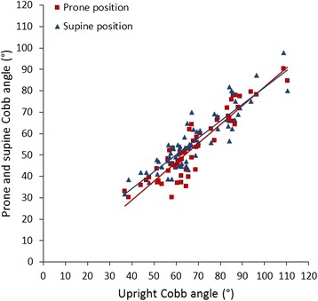
Results: Thoracic and lumbar Cobb angles correlated significantly between conventional radiographs (68° ± 15° and 44° ± 17°), prone CT (54° ± 15° and 33° ± 15°), and supine MRI (57° ± 14° and 35° ± 16°; ICC ≥0.96; P < 0.001). The thoracic and lumbar apical vertebral rotation showed a good correlation among three positions (upright, 22° ± 12° and 11° ± 13°; prone, 20° ± 9° and 8° ± 11°; supine, 16° ± 11° and 6° ± 14°; ICC ≥0.82; P < 0.001). The TK and LL correlated well among three different positions (TK 26° ± 11°, 22° ± 12°, and 17° ± 10°; P ≤ 0.004; LL 49° ± 12°, 45° ± 11°, and 44° ± 12°; P < 0.006; ICC 0.87 and 0.85).
Conclusions: Although there is a generalized underestimation of morphological parameters of the scoliotic deformity in the supine and prone positions as compared to the upright position, a significant correlation of these parameters is still evident among different body positions by different imaging modalities. Findings of this study suggest that severity of scoliotic deformity in AIS patients can be largely represented by different imaging modalities despite the difference in body positioning.
期刊介绍:
Cessation.Scoliosis and Spinal Disorders is an open access, multidisciplinary journal that encompasses all aspects of research on prevention, diagnosis, treatment, outcomes and cost-analyses of conservative and surgical management of all spinal deformities and disorders. Both clinical and basic science reports form the cornerstone of the journal in its endeavour to provide original, primary studies as well as narrative/systematic reviews and meta-analyses to the academic community and beyond. Scoliosis and Spinal Disorders aims to provide an integrated and balanced view of cutting-edge spine research to further enhance effective collaboration among clinical spine specialists and scientists, and to ultimately improve patient outcomes based on an evidence-based spine care approach.


 分享
分享
 求助内容:
求助内容: 应助结果提醒方式:
 扫码关注我们
