Carina Mari Aparici, Spencer C Behr, Youngho Seo, R Kate Kelley, Carlos Corvera, Kenneth T Gao, Rahul Aggarwal, Michael J Evans
{"title":"68ga -柠檬酸PET成像肝细胞癌:首次临床经验。","authors":"Carina Mari Aparici, Spencer C Behr, Youngho Seo, R Kate Kelley, Carlos Corvera, Kenneth T Gao, Rahul Aggarwal, Michael J Evans","doi":"10.1177/1536012117723256","DOIUrl":null,"url":null,"abstract":"<p><p>While cross-sectional imaging with computed tomography (CT) and magnetic resonance imaging is the primary method for diagnosing hepatocellular carcinoma (HCC), they provide little biological insight into this molecularly heterogeneous disease. Nuclear imaging tools that can detect molecular subsets of tumors could greatly improve diagnosis and management of HCC. To this end, we conducted a patient study to determine whether HCC can be resolved using <sup>68</sup>Ga-citrate positron emission tomography (PET). One patient with recurrent HCC was injected with 300 MBq of <sup>68</sup>Ga-citrate and imaged with PET/CT 249 minutes post injection. Four (28%) of 14 hepatic lesions were avid for <sup>68</sup>Ga-citrate. One extrahepatic lesion was not PET avid. The average maximum standardized uptake value (SUV<sub>max</sub>) for the lesions was 7.2 (range: 6.2-8.4), while the SUV<sub>max</sub> of the normal liver parenchyma was 4.7 and blood pool was 5.7. The avid lesions were not significantly larger than the quiescent lesions, and a prior contrast CT showed uniform enhancement among the lesions, suggesting that tumor signals are due to specific binding of the radiotracer to the transferrin receptor, rather than enhanced vascularity in the tumor microenvironment. Further studies are required in a larger patient cohort to verify the molecular basis of radiotracer uptake and the clinical utility of this tool.</p>","PeriodicalId":18855,"journal":{"name":"Molecular Imaging","volume":null,"pages":null},"PeriodicalIF":2.2000,"publicationDate":"2017-01-01","publicationTypes":"Journal Article","fieldsOfStudy":null,"isOpenAccess":false,"openAccessPdf":"https://sci-hub-pdf.com/10.1177/1536012117723256","citationCount":"7","resultStr":"{\"title\":\"Imaging Hepatocellular Carcinoma With <sup>68</sup>Ga-Citrate PET: First Clinical Experience.\",\"authors\":\"Carina Mari Aparici, Spencer C Behr, Youngho Seo, R Kate Kelley, Carlos Corvera, Kenneth T Gao, Rahul Aggarwal, Michael J Evans\",\"doi\":\"10.1177/1536012117723256\",\"DOIUrl\":null,\"url\":null,\"abstract\":\"<p><p>While cross-sectional imaging with computed tomography (CT) and magnetic resonance imaging is the primary method for diagnosing hepatocellular carcinoma (HCC), they provide little biological insight into this molecularly heterogeneous disease. Nuclear imaging tools that can detect molecular subsets of tumors could greatly improve diagnosis and management of HCC. To this end, we conducted a patient study to determine whether HCC can be resolved using <sup>68</sup>Ga-citrate positron emission tomography (PET). One patient with recurrent HCC was injected with 300 MBq of <sup>68</sup>Ga-citrate and imaged with PET/CT 249 minutes post injection. Four (28%) of 14 hepatic lesions were avid for <sup>68</sup>Ga-citrate. One extrahepatic lesion was not PET avid. The average maximum standardized uptake value (SUV<sub>max</sub>) for the lesions was 7.2 (range: 6.2-8.4), while the SUV<sub>max</sub> of the normal liver parenchyma was 4.7 and blood pool was 5.7. The avid lesions were not significantly larger than the quiescent lesions, and a prior contrast CT showed uniform enhancement among the lesions, suggesting that tumor signals are due to specific binding of the radiotracer to the transferrin receptor, rather than enhanced vascularity in the tumor microenvironment. Further studies are required in a larger patient cohort to verify the molecular basis of radiotracer uptake and the clinical utility of this tool.</p>\",\"PeriodicalId\":18855,\"journal\":{\"name\":\"Molecular Imaging\",\"volume\":null,\"pages\":null},\"PeriodicalIF\":2.2000,\"publicationDate\":\"2017-01-01\",\"publicationTypes\":\"Journal Article\",\"fieldsOfStudy\":null,\"isOpenAccess\":false,\"openAccessPdf\":\"https://sci-hub-pdf.com/10.1177/1536012117723256\",\"citationCount\":\"7\",\"resultStr\":null,\"platform\":\"Semanticscholar\",\"paperid\":null,\"PeriodicalName\":\"Molecular Imaging\",\"FirstCategoryId\":\"3\",\"ListUrlMain\":\"https://doi.org/10.1177/1536012117723256\",\"RegionNum\":4,\"RegionCategory\":\"医学\",\"ArticlePicture\":[],\"TitleCN\":null,\"AbstractTextCN\":null,\"PMCID\":null,\"EPubDate\":\"\",\"PubModel\":\"\",\"JCR\":\"Q3\",\"JCRName\":\"BIOCHEMICAL RESEARCH METHODS\",\"Score\":null,\"Total\":0}","platform":"Semanticscholar","paperid":null,"PeriodicalName":"Molecular Imaging","FirstCategoryId":"3","ListUrlMain":"https://doi.org/10.1177/1536012117723256","RegionNum":4,"RegionCategory":"医学","ArticlePicture":[],"TitleCN":null,"AbstractTextCN":null,"PMCID":null,"EPubDate":"","PubModel":"","JCR":"Q3","JCRName":"BIOCHEMICAL RESEARCH METHODS","Score":null,"Total":0}
Imaging Hepatocellular Carcinoma With 68Ga-Citrate PET: First Clinical Experience.
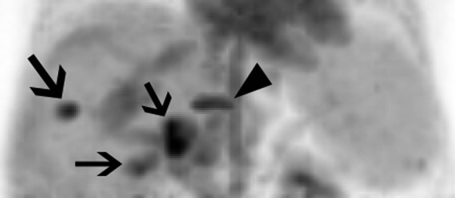
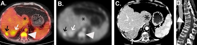
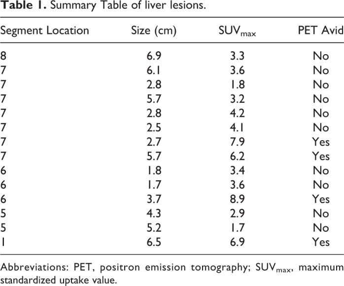
While cross-sectional imaging with computed tomography (CT) and magnetic resonance imaging is the primary method for diagnosing hepatocellular carcinoma (HCC), they provide little biological insight into this molecularly heterogeneous disease. Nuclear imaging tools that can detect molecular subsets of tumors could greatly improve diagnosis and management of HCC. To this end, we conducted a patient study to determine whether HCC can be resolved using 68Ga-citrate positron emission tomography (PET). One patient with recurrent HCC was injected with 300 MBq of 68Ga-citrate and imaged with PET/CT 249 minutes post injection. Four (28%) of 14 hepatic lesions were avid for 68Ga-citrate. One extrahepatic lesion was not PET avid. The average maximum standardized uptake value (SUVmax) for the lesions was 7.2 (range: 6.2-8.4), while the SUVmax of the normal liver parenchyma was 4.7 and blood pool was 5.7. The avid lesions were not significantly larger than the quiescent lesions, and a prior contrast CT showed uniform enhancement among the lesions, suggesting that tumor signals are due to specific binding of the radiotracer to the transferrin receptor, rather than enhanced vascularity in the tumor microenvironment. Further studies are required in a larger patient cohort to verify the molecular basis of radiotracer uptake and the clinical utility of this tool.
Molecular ImagingBiochemistry, Genetics and Molecular Biology-Biotechnology
自引率
3.60%
发文量
21
期刊介绍:
Molecular Imaging is a peer-reviewed, open access journal highlighting the breadth of molecular imaging research from basic science to preclinical studies to human applications. This serves both the scientific and clinical communities by disseminating novel results and concepts relevant to the biological study of normal and disease processes in both basic and translational studies ranging from mice to humans.


 分享
分享
 求助内容:
求助内容: 应助结果提醒方式:
 扫码关注我们
