{"title":"微波成像系统中基于EBG的微带贴片天线散射参数检测脑肿瘤。","authors":"Reefat Inum, Md Masud Rana, Kamrun Nahar Shushama, Md Anwarul Quader","doi":"10.1155/2018/8241438","DOIUrl":null,"url":null,"abstract":"<p><p>A microwave brain imaging system model is envisaged to detect and visualize tumor inside the human brain. A compact and efficient microstrip patch antenna is used in the imaging technique to transmit equivalent signal and receive backscattering signal from the stratified human head model. Electromagnetic band gap (EBG) structure is incorporated on the antenna ground plane to enhance the performance. Rectangular and circular EBG structures are proposed to investigate the antenna performance. Incorporation of circular EBG on the antenna ground plane provides an improvement of 22.77% in return loss, 5.84% in impedance bandwidth, and 16.53% in antenna gain with respect to the patch antenna with rectangular EBG. The simulation results obtained from CST are compared to those obtained from HFSS to validate the design. Specific absorption rate (SAR) of the modeled head tissue for the proposed antenna is determined. Different SAR values are compared with the established standard SAR limit to provide a safety regulation of the imaging system. A monostatic radar-based confocal microwave imaging algorithm is applied to generate the image of tumor inside a six-layer human head phantom model. <i>S</i>-parameter signals obtained from circular EBG loaded patch antenna in different scanning modes are utilized in the imaging algorithm to effectively produce a high-resolution image which reliably indicates the presence of tumor inside human brain.</p>","PeriodicalId":47063,"journal":{"name":"International Journal of Biomedical Imaging","volume":"2018 ","pages":"8241438"},"PeriodicalIF":1.3000,"publicationDate":"2018-02-12","publicationTypes":"Journal Article","fieldsOfStudy":null,"isOpenAccess":false,"openAccessPdf":"https://sci-hub-pdf.com/10.1155/2018/8241438","citationCount":"44","resultStr":"{\"title\":\"EBG Based Microstrip Patch Antenna for Brain Tumor Detection via Scattering Parameters in Microwave Imaging System.\",\"authors\":\"Reefat Inum, Md Masud Rana, Kamrun Nahar Shushama, Md Anwarul Quader\",\"doi\":\"10.1155/2018/8241438\",\"DOIUrl\":null,\"url\":null,\"abstract\":\"<p><p>A microwave brain imaging system model is envisaged to detect and visualize tumor inside the human brain. A compact and efficient microstrip patch antenna is used in the imaging technique to transmit equivalent signal and receive backscattering signal from the stratified human head model. Electromagnetic band gap (EBG) structure is incorporated on the antenna ground plane to enhance the performance. Rectangular and circular EBG structures are proposed to investigate the antenna performance. Incorporation of circular EBG on the antenna ground plane provides an improvement of 22.77% in return loss, 5.84% in impedance bandwidth, and 16.53% in antenna gain with respect to the patch antenna with rectangular EBG. The simulation results obtained from CST are compared to those obtained from HFSS to validate the design. Specific absorption rate (SAR) of the modeled head tissue for the proposed antenna is determined. Different SAR values are compared with the established standard SAR limit to provide a safety regulation of the imaging system. A monostatic radar-based confocal microwave imaging algorithm is applied to generate the image of tumor inside a six-layer human head phantom model. <i>S</i>-parameter signals obtained from circular EBG loaded patch antenna in different scanning modes are utilized in the imaging algorithm to effectively produce a high-resolution image which reliably indicates the presence of tumor inside human brain.</p>\",\"PeriodicalId\":47063,\"journal\":{\"name\":\"International Journal of Biomedical Imaging\",\"volume\":\"2018 \",\"pages\":\"8241438\"},\"PeriodicalIF\":1.3000,\"publicationDate\":\"2018-02-12\",\"publicationTypes\":\"Journal Article\",\"fieldsOfStudy\":null,\"isOpenAccess\":false,\"openAccessPdf\":\"https://sci-hub-pdf.com/10.1155/2018/8241438\",\"citationCount\":\"44\",\"resultStr\":null,\"platform\":\"Semanticscholar\",\"paperid\":null,\"PeriodicalName\":\"International Journal of Biomedical Imaging\",\"FirstCategoryId\":\"1085\",\"ListUrlMain\":\"https://doi.org/10.1155/2018/8241438\",\"RegionNum\":0,\"RegionCategory\":null,\"ArticlePicture\":[],\"TitleCN\":null,\"AbstractTextCN\":null,\"PMCID\":null,\"EPubDate\":\"2018/1/1 0:00:00\",\"PubModel\":\"eCollection\",\"JCR\":\"Q2\",\"JCRName\":\"ENGINEERING, BIOMEDICAL\",\"Score\":null,\"Total\":0}","platform":"Semanticscholar","paperid":null,"PeriodicalName":"International Journal of Biomedical Imaging","FirstCategoryId":"1085","ListUrlMain":"https://doi.org/10.1155/2018/8241438","RegionNum":0,"RegionCategory":null,"ArticlePicture":[],"TitleCN":null,"AbstractTextCN":null,"PMCID":null,"EPubDate":"2018/1/1 0:00:00","PubModel":"eCollection","JCR":"Q2","JCRName":"ENGINEERING, BIOMEDICAL","Score":null,"Total":0}
EBG Based Microstrip Patch Antenna for Brain Tumor Detection via Scattering Parameters in Microwave Imaging System.
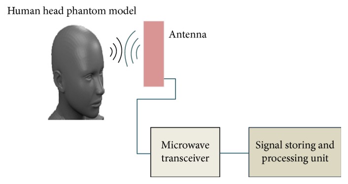
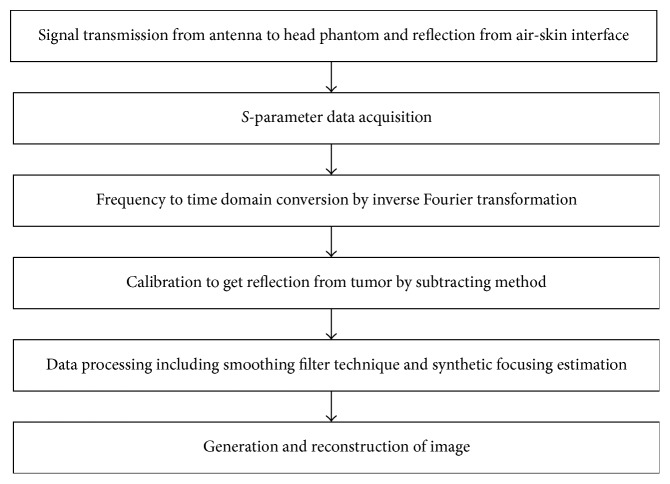
A microwave brain imaging system model is envisaged to detect and visualize tumor inside the human brain. A compact and efficient microstrip patch antenna is used in the imaging technique to transmit equivalent signal and receive backscattering signal from the stratified human head model. Electromagnetic band gap (EBG) structure is incorporated on the antenna ground plane to enhance the performance. Rectangular and circular EBG structures are proposed to investigate the antenna performance. Incorporation of circular EBG on the antenna ground plane provides an improvement of 22.77% in return loss, 5.84% in impedance bandwidth, and 16.53% in antenna gain with respect to the patch antenna with rectangular EBG. The simulation results obtained from CST are compared to those obtained from HFSS to validate the design. Specific absorption rate (SAR) of the modeled head tissue for the proposed antenna is determined. Different SAR values are compared with the established standard SAR limit to provide a safety regulation of the imaging system. A monostatic radar-based confocal microwave imaging algorithm is applied to generate the image of tumor inside a six-layer human head phantom model. S-parameter signals obtained from circular EBG loaded patch antenna in different scanning modes are utilized in the imaging algorithm to effectively produce a high-resolution image which reliably indicates the presence of tumor inside human brain.
期刊介绍:
The International Journal of Biomedical Imaging is managed by a board of editors comprising internationally renowned active researchers. The journal is freely accessible online and also offered for purchase in print format. It employs a web-based review system to ensure swift turnaround times while maintaining high standards. In addition to regular issues, special issues are organized by guest editors. The subject areas covered include (but are not limited to):
Digital radiography and tomosynthesis
X-ray computed tomography (CT)
Magnetic resonance imaging (MRI)
Single photon emission computed tomography (SPECT)
Positron emission tomography (PET)
Ultrasound imaging
Diffuse optical tomography, coherence, fluorescence, bioluminescence tomography, impedance tomography
Neutron imaging for biomedical applications
Magnetic and optical spectroscopy, and optical biopsy
Optical, electron, scanning tunneling/atomic force microscopy
Small animal imaging
Functional, cellular, and molecular imaging
Imaging assays for screening and molecular analysis
Microarray image analysis and bioinformatics
Emerging biomedical imaging techniques
Imaging modality fusion
Biomedical imaging instrumentation
Biomedical image processing, pattern recognition, and analysis
Biomedical image visualization, compression, transmission, and storage
Imaging and modeling related to systems biology and systems biomedicine
Applied mathematics, applied physics, and chemistry related to biomedical imaging
Grid-enabling technology for biomedical imaging and informatics


 分享
分享
 求助内容:
求助内容: 应助结果提醒方式:
 扫码关注我们
