Alberto Naoki Miyazaki, Luciana Andrade Silva, Pedro Doneux Santos, Guilherme do Val Sella, Leonardo Hideto Nagaya, Sergio Luiz Checchia
{"title":"肩前路不稳的Hill-Sachs损伤三维模型测量","authors":"Alberto Naoki Miyazaki, Luciana Andrade Silva, Pedro Doneux Santos, Guilherme do Val Sella, Leonardo Hideto Nagaya, Sergio Luiz Checchia","doi":"10.1016/j.rboe.2018.03.008","DOIUrl":null,"url":null,"abstract":"<div><h3>Objective</h3><p>To evaluate the reproducibility and repeatability of Hill–Sachs lesion measurement from computed tomography images, with computer software and tridimensional prototype.</p></div><div><h3>Methods</h3><p>Three-dimensional models were made from computed tomography images from 14 patients with anterior shoulder instability, using InVesalius 3.0® software. Hill–Sachs lesions were measured with Rhinocerus 5.0® software with pre-determined position. Mid-lateral distance, perpendicular to humeral shaft, cranial-caudal distance, parallel to humeral shaft, and the longitudinal distance of the lesion were measured. Using the Printer-ZP 310 three-dimensional printer, plaster models were made. To measure the Hill–Sachs lesion, a calibrated universal digital caliper was used in the same way as the software.</p></div><div><h3>Results</h3><p>There was intra-observer and inter-observer variability for measurement of the same model. Observers did not perform the measurements in a similar way, showing difficulty to use the method (<em>p</em> <!--><<!--> <!-->0.05). Using the software to measure the mid-lateral distance, as well as in the measurement with the caliper, the model type influenced the measurements for each of the observers, rendering the method invalid (<em>p</em> <!--><<!--> <!-->0.05).</p></div><div><h3>Conclusion</h3><p>There was no reproducibility and repeatability for Hill–Sachs lesion measurement between plaster models and software models.</p></div>","PeriodicalId":101095,"journal":{"name":"Revista Brasileira de Ortopedia (English Edition)","volume":"53 3","pages":"Pages 357-363"},"PeriodicalIF":0.0000,"publicationDate":"2018-05-01","publicationTypes":"Journal Article","fieldsOfStudy":null,"isOpenAccess":false,"openAccessPdf":"https://sci-hub-pdf.com/10.1016/j.rboe.2018.03.008","citationCount":"3","resultStr":"{\"title\":\"Hill–Sachs lesion measurement with tridimensional models in anterior shoulder instability\",\"authors\":\"Alberto Naoki Miyazaki, Luciana Andrade Silva, Pedro Doneux Santos, Guilherme do Val Sella, Leonardo Hideto Nagaya, Sergio Luiz Checchia\",\"doi\":\"10.1016/j.rboe.2018.03.008\",\"DOIUrl\":null,\"url\":null,\"abstract\":\"<div><h3>Objective</h3><p>To evaluate the reproducibility and repeatability of Hill–Sachs lesion measurement from computed tomography images, with computer software and tridimensional prototype.</p></div><div><h3>Methods</h3><p>Three-dimensional models were made from computed tomography images from 14 patients with anterior shoulder instability, using InVesalius 3.0® software. Hill–Sachs lesions were measured with Rhinocerus 5.0® software with pre-determined position. Mid-lateral distance, perpendicular to humeral shaft, cranial-caudal distance, parallel to humeral shaft, and the longitudinal distance of the lesion were measured. Using the Printer-ZP 310 three-dimensional printer, plaster models were made. To measure the Hill–Sachs lesion, a calibrated universal digital caliper was used in the same way as the software.</p></div><div><h3>Results</h3><p>There was intra-observer and inter-observer variability for measurement of the same model. Observers did not perform the measurements in a similar way, showing difficulty to use the method (<em>p</em> <!--><<!--> <!-->0.05). Using the software to measure the mid-lateral distance, as well as in the measurement with the caliper, the model type influenced the measurements for each of the observers, rendering the method invalid (<em>p</em> <!--><<!--> <!-->0.05).</p></div><div><h3>Conclusion</h3><p>There was no reproducibility and repeatability for Hill–Sachs lesion measurement between plaster models and software models.</p></div>\",\"PeriodicalId\":101095,\"journal\":{\"name\":\"Revista Brasileira de Ortopedia (English Edition)\",\"volume\":\"53 3\",\"pages\":\"Pages 357-363\"},\"PeriodicalIF\":0.0000,\"publicationDate\":\"2018-05-01\",\"publicationTypes\":\"Journal Article\",\"fieldsOfStudy\":null,\"isOpenAccess\":false,\"openAccessPdf\":\"https://sci-hub-pdf.com/10.1016/j.rboe.2018.03.008\",\"citationCount\":\"3\",\"resultStr\":null,\"platform\":\"Semanticscholar\",\"paperid\":null,\"PeriodicalName\":\"Revista Brasileira de Ortopedia (English Edition)\",\"FirstCategoryId\":\"1085\",\"ListUrlMain\":\"https://www.sciencedirect.com/science/article/pii/S2255497118300478\",\"RegionNum\":0,\"RegionCategory\":null,\"ArticlePicture\":[],\"TitleCN\":null,\"AbstractTextCN\":null,\"PMCID\":null,\"EPubDate\":\"\",\"PubModel\":\"\",\"JCR\":\"\",\"JCRName\":\"\",\"Score\":null,\"Total\":0}","platform":"Semanticscholar","paperid":null,"PeriodicalName":"Revista Brasileira de Ortopedia (English Edition)","FirstCategoryId":"1085","ListUrlMain":"https://www.sciencedirect.com/science/article/pii/S2255497118300478","RegionNum":0,"RegionCategory":null,"ArticlePicture":[],"TitleCN":null,"AbstractTextCN":null,"PMCID":null,"EPubDate":"","PubModel":"","JCR":"","JCRName":"","Score":null,"Total":0}
Hill–Sachs lesion measurement with tridimensional models in anterior shoulder instability
Objective
To evaluate the reproducibility and repeatability of Hill–Sachs lesion measurement from computed tomography images, with computer software and tridimensional prototype.
Methods
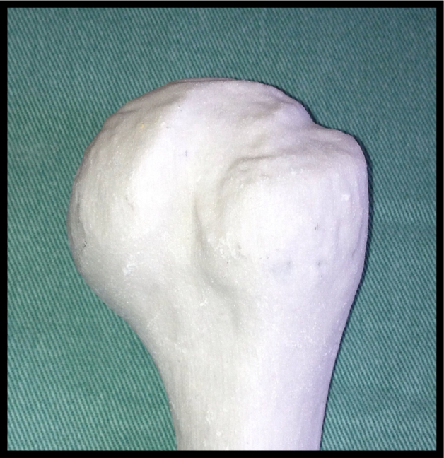
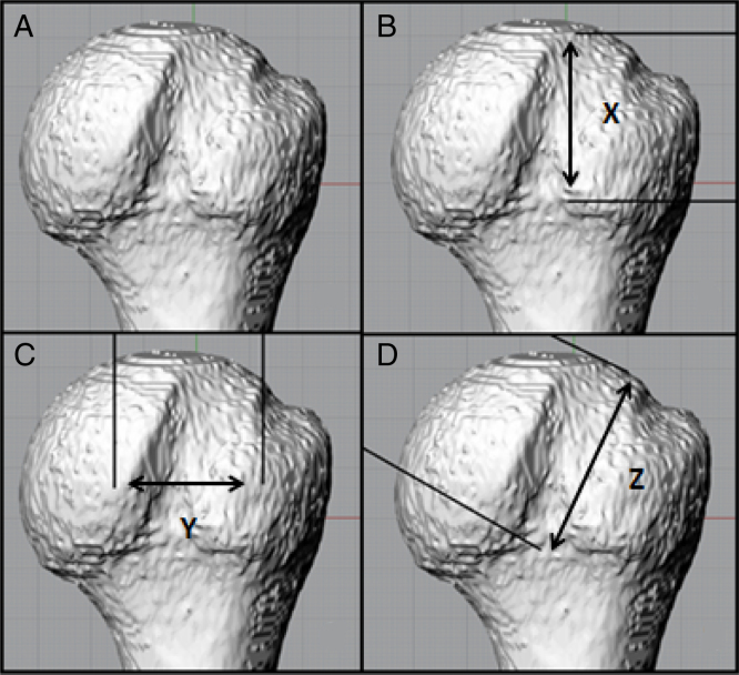
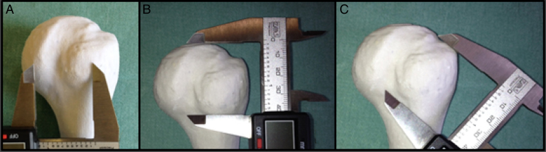
Three-dimensional models were made from computed tomography images from 14 patients with anterior shoulder instability, using InVesalius 3.0® software. Hill–Sachs lesions were measured with Rhinocerus 5.0® software with pre-determined position. Mid-lateral distance, perpendicular to humeral shaft, cranial-caudal distance, parallel to humeral shaft, and the longitudinal distance of the lesion were measured. Using the Printer-ZP 310 three-dimensional printer, plaster models were made. To measure the Hill–Sachs lesion, a calibrated universal digital caliper was used in the same way as the software.
Results
There was intra-observer and inter-observer variability for measurement of the same model. Observers did not perform the measurements in a similar way, showing difficulty to use the method (p < 0.05). Using the software to measure the mid-lateral distance, as well as in the measurement with the caliper, the model type influenced the measurements for each of the observers, rendering the method invalid (p < 0.05).
Conclusion
There was no reproducibility and repeatability for Hill–Sachs lesion measurement between plaster models and software models.


 分享
分享

 求助内容:
求助内容: 应助结果提醒方式:
 扫码关注我们
