Marieli Araujo Rossoni Marcioli , José Luis da Conceição Silva , Lucinéia de Fátima Chasko Ribeiro , Rose Meire Costa Brancalhão , Gladson Ricardo Flor Bertolini
{"title":"正中神经受压后神经活动Wistar大鼠神经营养因子表达及组织形态计量学评价","authors":"Marieli Araujo Rossoni Marcioli , José Luis da Conceição Silva , Lucinéia de Fátima Chasko Ribeiro , Rose Meire Costa Brancalhão , Gladson Ricardo Flor Bertolini","doi":"10.1016/j.rboe.2018.03.006","DOIUrl":null,"url":null,"abstract":"<div><h3>Objective</h3><p>To evaluate the neurotrophin mRNA expression and axon count in the median nerve of Wistar rats submitted to neural mobilization (NM) after nerve compression.</p></div><div><h3>Methods</h3><p>Eighteen animals were randomly divided into G1 (nerve compression only), G2 (NM for 1<!--> <!-->min), and G3 (NM for 3<!--> <!-->min). For NM, the animals were anesthetized and the right scapula received the mobilization, adapted as indicated for humans, on alternate days, from the third to the 13th postoperative (PO) day, totaling six days of therapy. On the 14th PO day, animals were anesthetized and euthanized. Fragments of the median nerve, distal to the compression procedure, were removed for histomorphometric analysis and expression of neurotrophins, nerve growth factor (NGF), and brain-derived neurotrophic factor (BDNF) by RT-PCR.</p></div><div><h3>Results</h3><p>Histomorphometric analysis revealed differences in the number of axons in the injured side, which was significantly lower in the injured limb nerve compared to the control limb, whereas the RT-PCR analysis showed no significant differences in the expression of NGF or BDNF.</p></div><div><h3>Conclusion</h3><p>NM treatment did not affect median nerve regeneration, which maintained normal recovery rates.</p></div>","PeriodicalId":101095,"journal":{"name":"Revista Brasileira de Ortopedia (English Edition)","volume":"53 3","pages":"Pages 276-280"},"PeriodicalIF":0.0000,"publicationDate":"2018-05-01","publicationTypes":"Journal Article","fieldsOfStudy":null,"isOpenAccess":false,"openAccessPdf":"https://sci-hub-pdf.com/10.1016/j.rboe.2018.03.006","citationCount":"3","resultStr":"{\"title\":\"Neurotrophin expression and histomorphometric evaluation in Wistar rats subjected to neural mobilization after compression of the median nerve\",\"authors\":\"Marieli Araujo Rossoni Marcioli , José Luis da Conceição Silva , Lucinéia de Fátima Chasko Ribeiro , Rose Meire Costa Brancalhão , Gladson Ricardo Flor Bertolini\",\"doi\":\"10.1016/j.rboe.2018.03.006\",\"DOIUrl\":null,\"url\":null,\"abstract\":\"<div><h3>Objective</h3><p>To evaluate the neurotrophin mRNA expression and axon count in the median nerve of Wistar rats submitted to neural mobilization (NM) after nerve compression.</p></div><div><h3>Methods</h3><p>Eighteen animals were randomly divided into G1 (nerve compression only), G2 (NM for 1<!--> <!-->min), and G3 (NM for 3<!--> <!-->min). For NM, the animals were anesthetized and the right scapula received the mobilization, adapted as indicated for humans, on alternate days, from the third to the 13th postoperative (PO) day, totaling six days of therapy. On the 14th PO day, animals were anesthetized and euthanized. Fragments of the median nerve, distal to the compression procedure, were removed for histomorphometric analysis and expression of neurotrophins, nerve growth factor (NGF), and brain-derived neurotrophic factor (BDNF) by RT-PCR.</p></div><div><h3>Results</h3><p>Histomorphometric analysis revealed differences in the number of axons in the injured side, which was significantly lower in the injured limb nerve compared to the control limb, whereas the RT-PCR analysis showed no significant differences in the expression of NGF or BDNF.</p></div><div><h3>Conclusion</h3><p>NM treatment did not affect median nerve regeneration, which maintained normal recovery rates.</p></div>\",\"PeriodicalId\":101095,\"journal\":{\"name\":\"Revista Brasileira de Ortopedia (English Edition)\",\"volume\":\"53 3\",\"pages\":\"Pages 276-280\"},\"PeriodicalIF\":0.0000,\"publicationDate\":\"2018-05-01\",\"publicationTypes\":\"Journal Article\",\"fieldsOfStudy\":null,\"isOpenAccess\":false,\"openAccessPdf\":\"https://sci-hub-pdf.com/10.1016/j.rboe.2018.03.006\",\"citationCount\":\"3\",\"resultStr\":null,\"platform\":\"Semanticscholar\",\"paperid\":null,\"PeriodicalName\":\"Revista Brasileira de Ortopedia (English Edition)\",\"FirstCategoryId\":\"1085\",\"ListUrlMain\":\"https://www.sciencedirect.com/science/article/pii/S2255497118300454\",\"RegionNum\":0,\"RegionCategory\":null,\"ArticlePicture\":[],\"TitleCN\":null,\"AbstractTextCN\":null,\"PMCID\":null,\"EPubDate\":\"2018/4/4 0:00:00\",\"PubModel\":\"Epub\",\"JCR\":\"\",\"JCRName\":\"\",\"Score\":null,\"Total\":0}","platform":"Semanticscholar","paperid":null,"PeriodicalName":"Revista Brasileira de Ortopedia (English Edition)","FirstCategoryId":"1085","ListUrlMain":"https://www.sciencedirect.com/science/article/pii/S2255497118300454","RegionNum":0,"RegionCategory":null,"ArticlePicture":[],"TitleCN":null,"AbstractTextCN":null,"PMCID":null,"EPubDate":"2018/4/4 0:00:00","PubModel":"Epub","JCR":"","JCRName":"","Score":null,"Total":0}
Neurotrophin expression and histomorphometric evaluation in Wistar rats subjected to neural mobilization after compression of the median nerve
Objective
To evaluate the neurotrophin mRNA expression and axon count in the median nerve of Wistar rats submitted to neural mobilization (NM) after nerve compression.
Methods
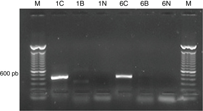
Eighteen animals were randomly divided into G1 (nerve compression only), G2 (NM for 1 min), and G3 (NM for 3 min). For NM, the animals were anesthetized and the right scapula received the mobilization, adapted as indicated for humans, on alternate days, from the third to the 13th postoperative (PO) day, totaling six days of therapy. On the 14th PO day, animals were anesthetized and euthanized. Fragments of the median nerve, distal to the compression procedure, were removed for histomorphometric analysis and expression of neurotrophins, nerve growth factor (NGF), and brain-derived neurotrophic factor (BDNF) by RT-PCR.
Results
Histomorphometric analysis revealed differences in the number of axons in the injured side, which was significantly lower in the injured limb nerve compared to the control limb, whereas the RT-PCR analysis showed no significant differences in the expression of NGF or BDNF.
Conclusion
NM treatment did not affect median nerve regeneration, which maintained normal recovery rates.


 分享
分享

 求助内容:
求助内容: 应助结果提醒方式:
 扫码关注我们
