{"title":"无瓣即刻种植及异种移植物材料填充颊骨间隙对颊骨水平的影响:一项随机临床试验。","authors":"Mojgan Paknejad, Solmaz Akbari, Hoori Aslroosta, Mehrdad Panjnoush, Samira Hajheidary","doi":"","DOIUrl":null,"url":null,"abstract":"<p><strong>Objectives: </strong>Following tooth extraction, soft and hard tissue alterations occur; Different factors can affect this process. The objective of this study was to determine the effect of gap filling on buccal alveolar crestal bone level after immediate implant placement after 4- to 6-month observation period.</p><p><strong>Materials and methods: </strong>This randomized clinical trial was performed on 20 patients (mean age of 38.8 years) requiring tooth extraction in a total of 27 areas in the anterior maxilla. The treatment strategy was as follows: atraumatic flapless tooth extraction, implant placement, insertion of a graft (test group) or no material (control group) between the implant and the socket wall, connection healing abutment placement and suturing the area. Clinical and cone beam computed tomographic examinations were performed before implant placement (baseline), 24 hours after surgery and 4-6 months (T2) after implant placement, to assess the buccal plate height (BH) and implant complications.</p><p><strong>Results: </strong>After 4 months of healing, a reduction in different bone measurements was noticed in the two groups. No statistically significant differences were assessed in bone height measurements between the test and control groups at different time points. The study demonstrated that immediate implantation resulted in 1.30 and 1.66 mm reduction in buccal bone plate in the test and control groups, respectively.</p><p><strong>Conclusions: </strong>The study demonstrated that immediate implantation in the extraction socket together with xenograft failed to prevent bone resorption.</p>","PeriodicalId":30286,"journal":{"name":"Journal of Dentistry of Tehran University of Medical Sciences","volume":"14 6","pages":"344-351"},"PeriodicalIF":0.0000,"publicationDate":"2017-11-01","publicationTypes":"Journal Article","fieldsOfStudy":null,"isOpenAccess":false,"openAccessPdf":"https://www.ncbi.nlm.nih.gov/pmc/articles/PMC6015591/pdf/","citationCount":"0","resultStr":"{\"title\":\"Effect of Flapless Immediate Implantation and Filling the Buccal Gap with Xenograft Material on the Buccal Bone Level: A Randomized Clinical Trial.\",\"authors\":\"Mojgan Paknejad, Solmaz Akbari, Hoori Aslroosta, Mehrdad Panjnoush, Samira Hajheidary\",\"doi\":\"\",\"DOIUrl\":null,\"url\":null,\"abstract\":\"<p><strong>Objectives: </strong>Following tooth extraction, soft and hard tissue alterations occur; Different factors can affect this process. The objective of this study was to determine the effect of gap filling on buccal alveolar crestal bone level after immediate implant placement after 4- to 6-month observation period.</p><p><strong>Materials and methods: </strong>This randomized clinical trial was performed on 20 patients (mean age of 38.8 years) requiring tooth extraction in a total of 27 areas in the anterior maxilla. The treatment strategy was as follows: atraumatic flapless tooth extraction, implant placement, insertion of a graft (test group) or no material (control group) between the implant and the socket wall, connection healing abutment placement and suturing the area. Clinical and cone beam computed tomographic examinations were performed before implant placement (baseline), 24 hours after surgery and 4-6 months (T2) after implant placement, to assess the buccal plate height (BH) and implant complications.</p><p><strong>Results: </strong>After 4 months of healing, a reduction in different bone measurements was noticed in the two groups. No statistically significant differences were assessed in bone height measurements between the test and control groups at different time points. The study demonstrated that immediate implantation resulted in 1.30 and 1.66 mm reduction in buccal bone plate in the test and control groups, respectively.</p><p><strong>Conclusions: </strong>The study demonstrated that immediate implantation in the extraction socket together with xenograft failed to prevent bone resorption.</p>\",\"PeriodicalId\":30286,\"journal\":{\"name\":\"Journal of Dentistry of Tehran University of Medical Sciences\",\"volume\":\"14 6\",\"pages\":\"344-351\"},\"PeriodicalIF\":0.0000,\"publicationDate\":\"2017-11-01\",\"publicationTypes\":\"Journal Article\",\"fieldsOfStudy\":null,\"isOpenAccess\":false,\"openAccessPdf\":\"https://www.ncbi.nlm.nih.gov/pmc/articles/PMC6015591/pdf/\",\"citationCount\":\"0\",\"resultStr\":null,\"platform\":\"Semanticscholar\",\"paperid\":null,\"PeriodicalName\":\"Journal of Dentistry of Tehran University of Medical Sciences\",\"FirstCategoryId\":\"1085\",\"ListUrlMain\":\"\",\"RegionNum\":0,\"RegionCategory\":null,\"ArticlePicture\":[],\"TitleCN\":null,\"AbstractTextCN\":null,\"PMCID\":null,\"EPubDate\":\"\",\"PubModel\":\"\",\"JCR\":\"\",\"JCRName\":\"\",\"Score\":null,\"Total\":0}","platform":"Semanticscholar","paperid":null,"PeriodicalName":"Journal of Dentistry of Tehran University of Medical Sciences","FirstCategoryId":"1085","ListUrlMain":"","RegionNum":0,"RegionCategory":null,"ArticlePicture":[],"TitleCN":null,"AbstractTextCN":null,"PMCID":null,"EPubDate":"","PubModel":"","JCR":"","JCRName":"","Score":null,"Total":0}
Effect of Flapless Immediate Implantation and Filling the Buccal Gap with Xenograft Material on the Buccal Bone Level: A Randomized Clinical Trial.
Objectives: Following tooth extraction, soft and hard tissue alterations occur; Different factors can affect this process. The objective of this study was to determine the effect of gap filling on buccal alveolar crestal bone level after immediate implant placement after 4- to 6-month observation period.
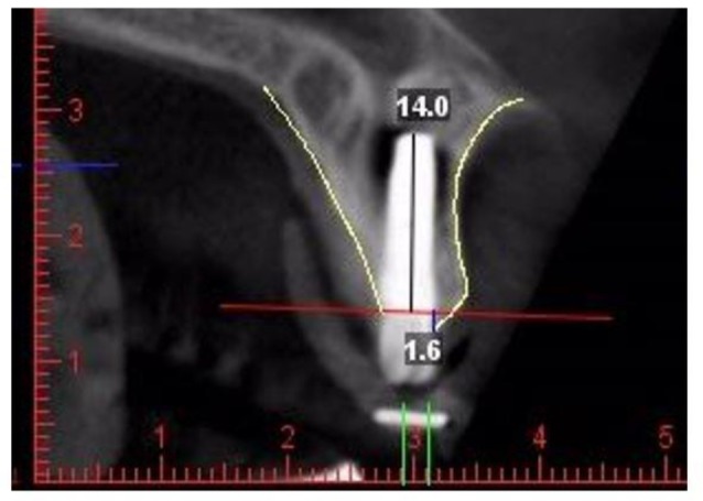
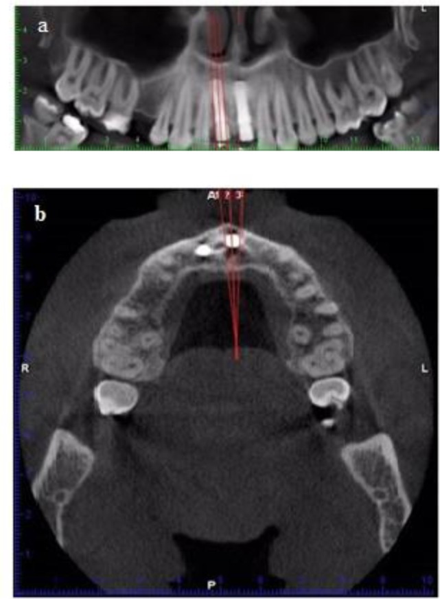
Materials and methods: This randomized clinical trial was performed on 20 patients (mean age of 38.8 years) requiring tooth extraction in a total of 27 areas in the anterior maxilla. The treatment strategy was as follows: atraumatic flapless tooth extraction, implant placement, insertion of a graft (test group) or no material (control group) between the implant and the socket wall, connection healing abutment placement and suturing the area. Clinical and cone beam computed tomographic examinations were performed before implant placement (baseline), 24 hours after surgery and 4-6 months (T2) after implant placement, to assess the buccal plate height (BH) and implant complications.
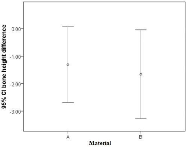
Results: After 4 months of healing, a reduction in different bone measurements was noticed in the two groups. No statistically significant differences were assessed in bone height measurements between the test and control groups at different time points. The study demonstrated that immediate implantation resulted in 1.30 and 1.66 mm reduction in buccal bone plate in the test and control groups, respectively.
Conclusions: The study demonstrated that immediate implantation in the extraction socket together with xenograft failed to prevent bone resorption.
 分享
分享


 求助内容:
求助内容: 应助结果提醒方式:
 扫码关注我们
