{"title":"应用术后图像估计颅骨切除术表面积。","authors":"Meng-Yin Ho, Wei-Lung Tseng, Furen Xiao","doi":"10.1155/2018/5237693","DOIUrl":null,"url":null,"abstract":"<p><p>Decompressive craniectomy (DC) is a neurosurgical procedure performed to relieve the intracranial pressure engendered by brain swelling. However, no easy and accurate method exists for determining the craniectomy surface area. In this study, we implemented and compared three methods of estimating the craniectomy surface area for evaluating the decompressive effort. We collected 118 sets of preoperative and postoperative brain computed tomography images from patients who underwent craniectomy procedures between April 2009 and April 2011. The surface area associated with each craniectomy was estimated using the marching cube and quasi-Monte Carlo methods. The surface area was also estimated using a simple AC method, in which the area is calculated by multiplying the craniectomy length (<i>A</i>) by its height (<i>C</i>). The estimated surface area ranged from 9.46 to 205.32 cm<sup>2</sup>, with a median of 134.80 cm<sup>2</sup>. The root-mean-square deviation (RMSD) between the marching cube and quasi-Monte Carlo methods was 7.53 cm<sup>2</sup>. Furthermore, the RMSD was 14.45 cm<sup>2</sup> between the marching cube and AC methods and 12.70 cm<sup>2</sup> between the quasi-Monte Carlo and AC methods. Paired <i>t</i>-tests indicated no statistically significant difference between these methods. The marching cube and quasi-Monte Carlo methods yield similar results. The results calculated using the AC method are also clinically acceptable for estimating the DC surface area. Our results can facilitate additional studies on the association of decompressive effort with the effect of craniectomy.</p>","PeriodicalId":47063,"journal":{"name":"International Journal of Biomedical Imaging","volume":"2018 ","pages":"5237693"},"PeriodicalIF":1.3000,"publicationDate":"2018-06-03","publicationTypes":"Journal Article","fieldsOfStudy":null,"isOpenAccess":false,"openAccessPdf":"https://sci-hub-pdf.com/10.1155/2018/5237693","citationCount":"7","resultStr":"{\"title\":\"Estimation of the Craniectomy Surface Area by Using Postoperative Images.\",\"authors\":\"Meng-Yin Ho, Wei-Lung Tseng, Furen Xiao\",\"doi\":\"10.1155/2018/5237693\",\"DOIUrl\":null,\"url\":null,\"abstract\":\"<p><p>Decompressive craniectomy (DC) is a neurosurgical procedure performed to relieve the intracranial pressure engendered by brain swelling. However, no easy and accurate method exists for determining the craniectomy surface area. In this study, we implemented and compared three methods of estimating the craniectomy surface area for evaluating the decompressive effort. We collected 118 sets of preoperative and postoperative brain computed tomography images from patients who underwent craniectomy procedures between April 2009 and April 2011. The surface area associated with each craniectomy was estimated using the marching cube and quasi-Monte Carlo methods. The surface area was also estimated using a simple AC method, in which the area is calculated by multiplying the craniectomy length (<i>A</i>) by its height (<i>C</i>). The estimated surface area ranged from 9.46 to 205.32 cm<sup>2</sup>, with a median of 134.80 cm<sup>2</sup>. The root-mean-square deviation (RMSD) between the marching cube and quasi-Monte Carlo methods was 7.53 cm<sup>2</sup>. Furthermore, the RMSD was 14.45 cm<sup>2</sup> between the marching cube and AC methods and 12.70 cm<sup>2</sup> between the quasi-Monte Carlo and AC methods. Paired <i>t</i>-tests indicated no statistically significant difference between these methods. The marching cube and quasi-Monte Carlo methods yield similar results. The results calculated using the AC method are also clinically acceptable for estimating the DC surface area. Our results can facilitate additional studies on the association of decompressive effort with the effect of craniectomy.</p>\",\"PeriodicalId\":47063,\"journal\":{\"name\":\"International Journal of Biomedical Imaging\",\"volume\":\"2018 \",\"pages\":\"5237693\"},\"PeriodicalIF\":1.3000,\"publicationDate\":\"2018-06-03\",\"publicationTypes\":\"Journal Article\",\"fieldsOfStudy\":null,\"isOpenAccess\":false,\"openAccessPdf\":\"https://sci-hub-pdf.com/10.1155/2018/5237693\",\"citationCount\":\"7\",\"resultStr\":null,\"platform\":\"Semanticscholar\",\"paperid\":null,\"PeriodicalName\":\"International Journal of Biomedical Imaging\",\"FirstCategoryId\":\"1085\",\"ListUrlMain\":\"https://doi.org/10.1155/2018/5237693\",\"RegionNum\":0,\"RegionCategory\":null,\"ArticlePicture\":[],\"TitleCN\":null,\"AbstractTextCN\":null,\"PMCID\":null,\"EPubDate\":\"2018/1/1 0:00:00\",\"PubModel\":\"eCollection\",\"JCR\":\"Q2\",\"JCRName\":\"ENGINEERING, BIOMEDICAL\",\"Score\":null,\"Total\":0}","platform":"Semanticscholar","paperid":null,"PeriodicalName":"International Journal of Biomedical Imaging","FirstCategoryId":"1085","ListUrlMain":"https://doi.org/10.1155/2018/5237693","RegionNum":0,"RegionCategory":null,"ArticlePicture":[],"TitleCN":null,"AbstractTextCN":null,"PMCID":null,"EPubDate":"2018/1/1 0:00:00","PubModel":"eCollection","JCR":"Q2","JCRName":"ENGINEERING, BIOMEDICAL","Score":null,"Total":0}
Estimation of the Craniectomy Surface Area by Using Postoperative Images.
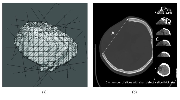
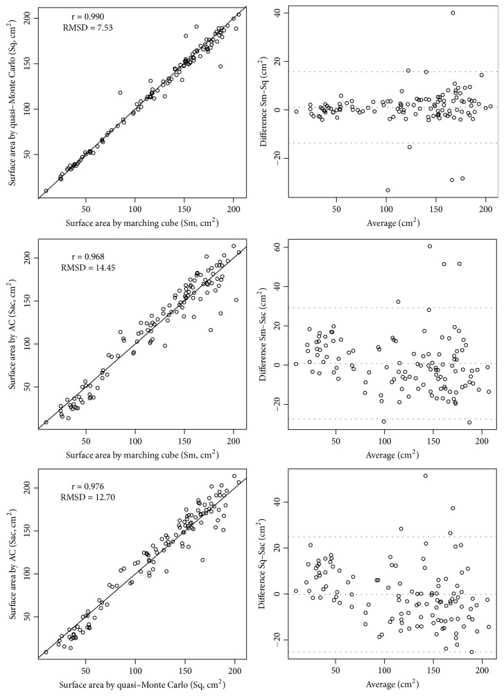
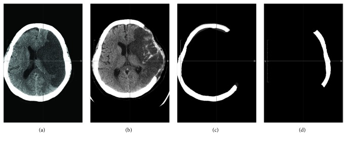
Decompressive craniectomy (DC) is a neurosurgical procedure performed to relieve the intracranial pressure engendered by brain swelling. However, no easy and accurate method exists for determining the craniectomy surface area. In this study, we implemented and compared three methods of estimating the craniectomy surface area for evaluating the decompressive effort. We collected 118 sets of preoperative and postoperative brain computed tomography images from patients who underwent craniectomy procedures between April 2009 and April 2011. The surface area associated with each craniectomy was estimated using the marching cube and quasi-Monte Carlo methods. The surface area was also estimated using a simple AC method, in which the area is calculated by multiplying the craniectomy length (A) by its height (C). The estimated surface area ranged from 9.46 to 205.32 cm2, with a median of 134.80 cm2. The root-mean-square deviation (RMSD) between the marching cube and quasi-Monte Carlo methods was 7.53 cm2. Furthermore, the RMSD was 14.45 cm2 between the marching cube and AC methods and 12.70 cm2 between the quasi-Monte Carlo and AC methods. Paired t-tests indicated no statistically significant difference between these methods. The marching cube and quasi-Monte Carlo methods yield similar results. The results calculated using the AC method are also clinically acceptable for estimating the DC surface area. Our results can facilitate additional studies on the association of decompressive effort with the effect of craniectomy.
期刊介绍:
The International Journal of Biomedical Imaging is managed by a board of editors comprising internationally renowned active researchers. The journal is freely accessible online and also offered for purchase in print format. It employs a web-based review system to ensure swift turnaround times while maintaining high standards. In addition to regular issues, special issues are organized by guest editors. The subject areas covered include (but are not limited to):
Digital radiography and tomosynthesis
X-ray computed tomography (CT)
Magnetic resonance imaging (MRI)
Single photon emission computed tomography (SPECT)
Positron emission tomography (PET)
Ultrasound imaging
Diffuse optical tomography, coherence, fluorescence, bioluminescence tomography, impedance tomography
Neutron imaging for biomedical applications
Magnetic and optical spectroscopy, and optical biopsy
Optical, electron, scanning tunneling/atomic force microscopy
Small animal imaging
Functional, cellular, and molecular imaging
Imaging assays for screening and molecular analysis
Microarray image analysis and bioinformatics
Emerging biomedical imaging techniques
Imaging modality fusion
Biomedical imaging instrumentation
Biomedical image processing, pattern recognition, and analysis
Biomedical image visualization, compression, transmission, and storage
Imaging and modeling related to systems biology and systems biomedicine
Applied mathematics, applied physics, and chemistry related to biomedical imaging
Grid-enabling technology for biomedical imaging and informatics


 分享
分享
 求助内容:
求助内容: 应助结果提醒方式:
 扫码关注我们
