Maryam Khoroush, Fatemeh Keshani, Mehdi Esmaeili, Moeen Hosseini Shirazi
{"title":"受龋影响的牙本质壁颈部修复体边缘完整性:止血剂污染的影响。","authors":"Maryam Khoroush, Fatemeh Keshani, Mehdi Esmaeili, Moeen Hosseini Shirazi","doi":"","DOIUrl":null,"url":null,"abstract":"<p><strong>Objectives: </strong>The aim of this study was to compare the microleakage in normal and caries-affected dentin (CAD) and to investigate the effect of three hemostatic agents on the microleakage of Class V composite resin restorations in CAD.</p><p><strong>Materials and methods: </strong>Ninety-six Class V non-beveled cavities were prepared in 48 third molars at 1 mm below the cementoenamel junction (CEJ) in the cervical margin with the occlusogingival size of 2 mm, mesiodistal dimension of 3 mm, and a depth of 1.5 mm. Next, the teeth were divided into 8 groups (n=12): G1-4 included normal dentin (N) substrate, while G5-8 were exposed to mineralization/demineralization cycles to produce CAD substrate. Groups 1 and 5 were the controls. ViscoStat was used in groups 2 and 6, ViscoStat Clear was used in groups 3 and 7, while trichloroacetic acid (TCA) was used in groups 4 and 8. The cavities were restored with composite resin. The samples were sectioned after thermocycling and immersion in 2% fuchsin for 24 hours. The degree of dye penetration was evaluated under a stereomicroscope at 40× magnification. Data were evaluated using Kruskal-Wallis and Mann-U-Whitney tests in SPSS 15 software (α=0.05).</p><p><strong>Results: </strong>Significant differences were recorded on the mean microleakage of different groups (P=0.047). There was a significant difference in the mean dentinal microleakage between N and CAD groups (P=0.014). The dentinal microleakage in group 3 was significantly higher than that in groups 4 to 8.</p><p><strong>Conclusions: </strong>According to the results, CAD showed less microleakage in comparison with intact dentin. ViscoStat Clear caused a greater microleakage than did ViscoStat or TCA.</p>","PeriodicalId":30286,"journal":{"name":"Journal of Dentistry of Tehran University of Medical Sciences","volume":"15 4","pages":"214-221"},"PeriodicalIF":0.0000,"publicationDate":"2018-07-01","publicationTypes":"Journal Article","fieldsOfStudy":null,"isOpenAccess":false,"openAccessPdf":"https://www.ncbi.nlm.nih.gov/pmc/articles/PMC6218462/pdf/","citationCount":"0","resultStr":"{\"title\":\"Marginal Integrity of Cervical Restorations with Caries-Affected Dentinal Walls: Effect of Contamination with Hemostatic Agents.\",\"authors\":\"Maryam Khoroush, Fatemeh Keshani, Mehdi Esmaeili, Moeen Hosseini Shirazi\",\"doi\":\"\",\"DOIUrl\":null,\"url\":null,\"abstract\":\"<p><strong>Objectives: </strong>The aim of this study was to compare the microleakage in normal and caries-affected dentin (CAD) and to investigate the effect of three hemostatic agents on the microleakage of Class V composite resin restorations in CAD.</p><p><strong>Materials and methods: </strong>Ninety-six Class V non-beveled cavities were prepared in 48 third molars at 1 mm below the cementoenamel junction (CEJ) in the cervical margin with the occlusogingival size of 2 mm, mesiodistal dimension of 3 mm, and a depth of 1.5 mm. Next, the teeth were divided into 8 groups (n=12): G1-4 included normal dentin (N) substrate, while G5-8 were exposed to mineralization/demineralization cycles to produce CAD substrate. Groups 1 and 5 were the controls. ViscoStat was used in groups 2 and 6, ViscoStat Clear was used in groups 3 and 7, while trichloroacetic acid (TCA) was used in groups 4 and 8. The cavities were restored with composite resin. The samples were sectioned after thermocycling and immersion in 2% fuchsin for 24 hours. The degree of dye penetration was evaluated under a stereomicroscope at 40× magnification. Data were evaluated using Kruskal-Wallis and Mann-U-Whitney tests in SPSS 15 software (α=0.05).</p><p><strong>Results: </strong>Significant differences were recorded on the mean microleakage of different groups (P=0.047). There was a significant difference in the mean dentinal microleakage between N and CAD groups (P=0.014). The dentinal microleakage in group 3 was significantly higher than that in groups 4 to 8.</p><p><strong>Conclusions: </strong>According to the results, CAD showed less microleakage in comparison with intact dentin. ViscoStat Clear caused a greater microleakage than did ViscoStat or TCA.</p>\",\"PeriodicalId\":30286,\"journal\":{\"name\":\"Journal of Dentistry of Tehran University of Medical Sciences\",\"volume\":\"15 4\",\"pages\":\"214-221\"},\"PeriodicalIF\":0.0000,\"publicationDate\":\"2018-07-01\",\"publicationTypes\":\"Journal Article\",\"fieldsOfStudy\":null,\"isOpenAccess\":false,\"openAccessPdf\":\"https://www.ncbi.nlm.nih.gov/pmc/articles/PMC6218462/pdf/\",\"citationCount\":\"0\",\"resultStr\":null,\"platform\":\"Semanticscholar\",\"paperid\":null,\"PeriodicalName\":\"Journal of Dentistry of Tehran University of Medical Sciences\",\"FirstCategoryId\":\"1085\",\"ListUrlMain\":\"\",\"RegionNum\":0,\"RegionCategory\":null,\"ArticlePicture\":[],\"TitleCN\":null,\"AbstractTextCN\":null,\"PMCID\":null,\"EPubDate\":\"\",\"PubModel\":\"\",\"JCR\":\"\",\"JCRName\":\"\",\"Score\":null,\"Total\":0}","platform":"Semanticscholar","paperid":null,"PeriodicalName":"Journal of Dentistry of Tehran University of Medical Sciences","FirstCategoryId":"1085","ListUrlMain":"","RegionNum":0,"RegionCategory":null,"ArticlePicture":[],"TitleCN":null,"AbstractTextCN":null,"PMCID":null,"EPubDate":"","PubModel":"","JCR":"","JCRName":"","Score":null,"Total":0}
Marginal Integrity of Cervical Restorations with Caries-Affected Dentinal Walls: Effect of Contamination with Hemostatic Agents.
Objectives: The aim of this study was to compare the microleakage in normal and caries-affected dentin (CAD) and to investigate the effect of three hemostatic agents on the microleakage of Class V composite resin restorations in CAD.
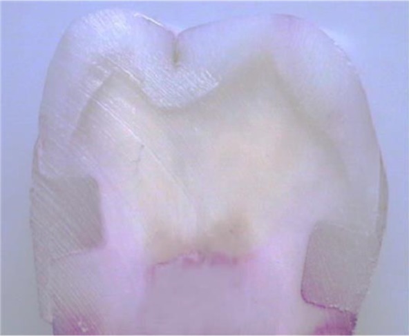
Materials and methods: Ninety-six Class V non-beveled cavities were prepared in 48 third molars at 1 mm below the cementoenamel junction (CEJ) in the cervical margin with the occlusogingival size of 2 mm, mesiodistal dimension of 3 mm, and a depth of 1.5 mm. Next, the teeth were divided into 8 groups (n=12): G1-4 included normal dentin (N) substrate, while G5-8 were exposed to mineralization/demineralization cycles to produce CAD substrate. Groups 1 and 5 were the controls. ViscoStat was used in groups 2 and 6, ViscoStat Clear was used in groups 3 and 7, while trichloroacetic acid (TCA) was used in groups 4 and 8. The cavities were restored with composite resin. The samples were sectioned after thermocycling and immersion in 2% fuchsin for 24 hours. The degree of dye penetration was evaluated under a stereomicroscope at 40× magnification. Data were evaluated using Kruskal-Wallis and Mann-U-Whitney tests in SPSS 15 software (α=0.05).
Results: Significant differences were recorded on the mean microleakage of different groups (P=0.047). There was a significant difference in the mean dentinal microleakage between N and CAD groups (P=0.014). The dentinal microleakage in group 3 was significantly higher than that in groups 4 to 8.
Conclusions: According to the results, CAD showed less microleakage in comparison with intact dentin. ViscoStat Clear caused a greater microleakage than did ViscoStat or TCA.

 分享
分享


 求助内容:
求助内容: 应助结果提醒方式:
 扫码关注我们
