Igor Rabelo de França, Daniela Meneses-Santos, Gabriela Virginia Moreira, Fábio Bessa Lima, Carla Roberta de Oliveira Carvalho, Anderson Carlos Marçal
{"title":"地塞米松治疗大鼠咬肌胰岛素信号通路的研究。","authors":"Igor Rabelo de França, Daniela Meneses-Santos, Gabriela Virginia Moreira, Fábio Bessa Lima, Carla Roberta de Oliveira Carvalho, Anderson Carlos Marçal","doi":"10.1556/1646.10.2018.44","DOIUrl":null,"url":null,"abstract":"<p><strong>Background and aims: </strong>The treatment with glucocorticoids may induce molecular changes in the level and/or degree of phosphorylation of proteins located downstream of the insulin receptor/insulin-like growth factor receptor (IR/IGF1R) in many tissues. However, few studies have investigated the intracellular insulin pathway in the masseter muscle. Therefore, this study aimed to analyze the IR/IGF1R signaling pathway in the masseter muscle of rats treated with dexamethasone.</p><p><strong>Materials and methods: </strong>Male Wistar rats were divided into two groups: control group (intraperitoneally injected with 0.9% NaCl solution) and dexamethasone group [intraperitoneally injected with 1 mg/kg (bw) dexamethasone solution] for 10 consecutive days. Sections of the masseter muscle were removed at time zero and after the infusion of regular insulin into the portal vein.</p><p><strong>Results: </strong>Dexamethasone administration induces body weight loss without changing masseter muscle weight and reduces the expression of total IR and PI3K proteins; total levels of IRS1, Akt, and ERK1 remain unchanged between groups. The degree of phosphorylation/activity of IRS1 after insulin stimulus increased only in the control group; degree of phosphorylation of Akt increased in both groups, but this increase was attenuated in the dexamethasone group.</p><p><strong>Discussion and conclusion: </strong>The degree of phosphorylation/activity in the masseter muscle is different from that in other muscle territories.</p>","PeriodicalId":45181,"journal":{"name":"Interventional Medicine and Applied Science","volume":"10 4","pages":"226-232"},"PeriodicalIF":0.0000,"publicationDate":"2018-12-01","publicationTypes":"Journal Article","fieldsOfStudy":null,"isOpenAccess":false,"openAccessPdf":"https://sci-hub-pdf.com/10.1556/1646.10.2018.44","citationCount":"3","resultStr":"{\"title\":\"Insulin signaling pathway in the masseter muscle of dexamethasone-treated rats.\",\"authors\":\"Igor Rabelo de França, Daniela Meneses-Santos, Gabriela Virginia Moreira, Fábio Bessa Lima, Carla Roberta de Oliveira Carvalho, Anderson Carlos Marçal\",\"doi\":\"10.1556/1646.10.2018.44\",\"DOIUrl\":null,\"url\":null,\"abstract\":\"<p><strong>Background and aims: </strong>The treatment with glucocorticoids may induce molecular changes in the level and/or degree of phosphorylation of proteins located downstream of the insulin receptor/insulin-like growth factor receptor (IR/IGF1R) in many tissues. However, few studies have investigated the intracellular insulin pathway in the masseter muscle. Therefore, this study aimed to analyze the IR/IGF1R signaling pathway in the masseter muscle of rats treated with dexamethasone.</p><p><strong>Materials and methods: </strong>Male Wistar rats were divided into two groups: control group (intraperitoneally injected with 0.9% NaCl solution) and dexamethasone group [intraperitoneally injected with 1 mg/kg (bw) dexamethasone solution] for 10 consecutive days. Sections of the masseter muscle were removed at time zero and after the infusion of regular insulin into the portal vein.</p><p><strong>Results: </strong>Dexamethasone administration induces body weight loss without changing masseter muscle weight and reduces the expression of total IR and PI3K proteins; total levels of IRS1, Akt, and ERK1 remain unchanged between groups. The degree of phosphorylation/activity of IRS1 after insulin stimulus increased only in the control group; degree of phosphorylation of Akt increased in both groups, but this increase was attenuated in the dexamethasone group.</p><p><strong>Discussion and conclusion: </strong>The degree of phosphorylation/activity in the masseter muscle is different from that in other muscle territories.</p>\",\"PeriodicalId\":45181,\"journal\":{\"name\":\"Interventional Medicine and Applied Science\",\"volume\":\"10 4\",\"pages\":\"226-232\"},\"PeriodicalIF\":0.0000,\"publicationDate\":\"2018-12-01\",\"publicationTypes\":\"Journal Article\",\"fieldsOfStudy\":null,\"isOpenAccess\":false,\"openAccessPdf\":\"https://sci-hub-pdf.com/10.1556/1646.10.2018.44\",\"citationCount\":\"3\",\"resultStr\":null,\"platform\":\"Semanticscholar\",\"paperid\":null,\"PeriodicalName\":\"Interventional Medicine and Applied Science\",\"FirstCategoryId\":\"1085\",\"ListUrlMain\":\"https://doi.org/10.1556/1646.10.2018.44\",\"RegionNum\":0,\"RegionCategory\":null,\"ArticlePicture\":[],\"TitleCN\":null,\"AbstractTextCN\":null,\"PMCID\":null,\"EPubDate\":\"\",\"PubModel\":\"\",\"JCR\":\"Q2\",\"JCRName\":\"Medicine\",\"Score\":null,\"Total\":0}","platform":"Semanticscholar","paperid":null,"PeriodicalName":"Interventional Medicine and Applied Science","FirstCategoryId":"1085","ListUrlMain":"https://doi.org/10.1556/1646.10.2018.44","RegionNum":0,"RegionCategory":null,"ArticlePicture":[],"TitleCN":null,"AbstractTextCN":null,"PMCID":null,"EPubDate":"","PubModel":"","JCR":"Q2","JCRName":"Medicine","Score":null,"Total":0}
Insulin signaling pathway in the masseter muscle of dexamethasone-treated rats.
Background and aims: The treatment with glucocorticoids may induce molecular changes in the level and/or degree of phosphorylation of proteins located downstream of the insulin receptor/insulin-like growth factor receptor (IR/IGF1R) in many tissues. However, few studies have investigated the intracellular insulin pathway in the masseter muscle. Therefore, this study aimed to analyze the IR/IGF1R signaling pathway in the masseter muscle of rats treated with dexamethasone.
Materials and methods: Male Wistar rats were divided into two groups: control group (intraperitoneally injected with 0.9% NaCl solution) and dexamethasone group [intraperitoneally injected with 1 mg/kg (bw) dexamethasone solution] for 10 consecutive days. Sections of the masseter muscle were removed at time zero and after the infusion of regular insulin into the portal vein.
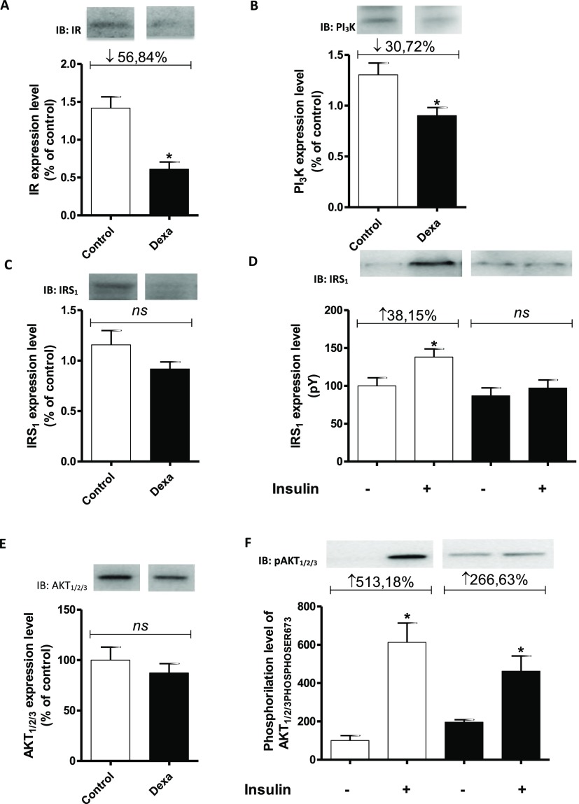
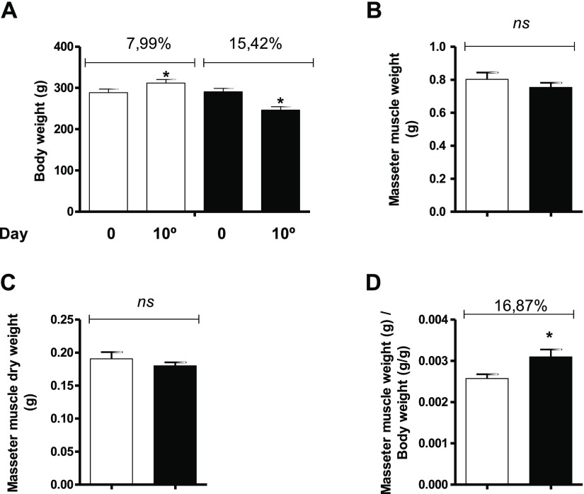
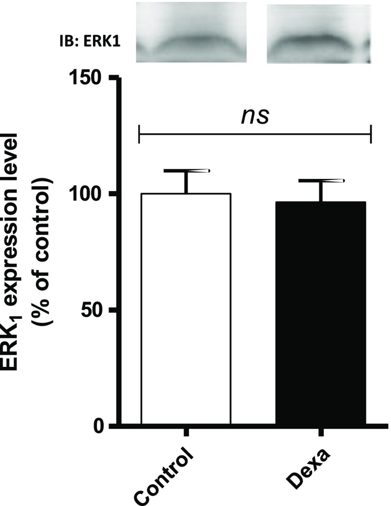
Results: Dexamethasone administration induces body weight loss without changing masseter muscle weight and reduces the expression of total IR and PI3K proteins; total levels of IRS1, Akt, and ERK1 remain unchanged between groups. The degree of phosphorylation/activity of IRS1 after insulin stimulus increased only in the control group; degree of phosphorylation of Akt increased in both groups, but this increase was attenuated in the dexamethasone group.
Discussion and conclusion: The degree of phosphorylation/activity in the masseter muscle is different from that in other muscle territories.


 分享
分享
 求助内容:
求助内容: 应助结果提醒方式:
 扫码关注我们
