{"title":"足菌肿骨组织常规脱钙与微波辅助脱钙的比较。","authors":"Magdi Mansour Salih","doi":"10.1155/2020/6561980","DOIUrl":null,"url":null,"abstract":"<p><p>Mycetoma is a lifelong granulomatous disease of subcutaneous tissues and bones. Histopathology is a substantiated indicative method based on the assumption of a definitive diagnosis of mycetoma. It requires efficient processing of tissues including bone decalcification. The decalcification process must ensure complete removal of calcium and also a proper preservation of tissue and microorganisms' staining ability. <i>Objectives</i>. To compare the conventional method used in decalcification with the microwave method using different decalcification solutions. Different characteristics were tested, including the speed of decalcification and morphological and fungal preservation in bone tissue affected with mycetoma. <i>Materials and Methods</i>. Three decalcification solutions were employed to remove calcium from 50 bone tissue samples affected with mycetoma, including 10% neutral buffered EDTA (pH 7.4), 5% nitric acid, and 5% hydrochloric acid. Conventional and microwave methods were used. Haematoxylin-eosin (HE) stain, Gridley's stain, and Grocott hexamine-silver stain were employed to evaluate the bone and fungi morphologies. <i>Results</i>. The decalcification time of the conventional method compared with the microwave method with 10% EDTA (pH 7.4) took 120 hours and 29 hours, while 5% hydrochloric acid and 5% nitric acid took 8 hours and 3 hours, separately. Also, 10% EDTA is the best decalcifying agent for HE staining and fungal stains. 5% hydrochloric acid and 5% nitric acid can be used for fungal staining. <i>Conclusion</i>. The current study investigated the effects of different decalcifying agents as well as two decalcification procedures on the preservation of the bone structure and fungal staining, which will help to develop suitable protocols for the analyses of the bone tissue affected with mycetoma infection.</p>","PeriodicalId":8826,"journal":{"name":"Biochemistry Research International","volume":"2020 ","pages":"6561980"},"PeriodicalIF":2.9000,"publicationDate":"2020-08-01","publicationTypes":"Journal Article","fieldsOfStudy":null,"isOpenAccess":false,"openAccessPdf":"https://sci-hub-pdf.com/10.1155/2020/6561980","citationCount":"4","resultStr":"{\"title\":\"Comparison between Conventional Decalcification and a Microwave-Assisted Method in Bone Tissue Affected with Mycetoma.\",\"authors\":\"Magdi Mansour Salih\",\"doi\":\"10.1155/2020/6561980\",\"DOIUrl\":null,\"url\":null,\"abstract\":\"<p><p>Mycetoma is a lifelong granulomatous disease of subcutaneous tissues and bones. Histopathology is a substantiated indicative method based on the assumption of a definitive diagnosis of mycetoma. It requires efficient processing of tissues including bone decalcification. The decalcification process must ensure complete removal of calcium and also a proper preservation of tissue and microorganisms' staining ability. <i>Objectives</i>. To compare the conventional method used in decalcification with the microwave method using different decalcification solutions. Different characteristics were tested, including the speed of decalcification and morphological and fungal preservation in bone tissue affected with mycetoma. <i>Materials and Methods</i>. Three decalcification solutions were employed to remove calcium from 50 bone tissue samples affected with mycetoma, including 10% neutral buffered EDTA (pH 7.4), 5% nitric acid, and 5% hydrochloric acid. Conventional and microwave methods were used. Haematoxylin-eosin (HE) stain, Gridley's stain, and Grocott hexamine-silver stain were employed to evaluate the bone and fungi morphologies. <i>Results</i>. The decalcification time of the conventional method compared with the microwave method with 10% EDTA (pH 7.4) took 120 hours and 29 hours, while 5% hydrochloric acid and 5% nitric acid took 8 hours and 3 hours, separately. Also, 10% EDTA is the best decalcifying agent for HE staining and fungal stains. 5% hydrochloric acid and 5% nitric acid can be used for fungal staining. <i>Conclusion</i>. The current study investigated the effects of different decalcifying agents as well as two decalcification procedures on the preservation of the bone structure and fungal staining, which will help to develop suitable protocols for the analyses of the bone tissue affected with mycetoma infection.</p>\",\"PeriodicalId\":8826,\"journal\":{\"name\":\"Biochemistry Research International\",\"volume\":\"2020 \",\"pages\":\"6561980\"},\"PeriodicalIF\":2.9000,\"publicationDate\":\"2020-08-01\",\"publicationTypes\":\"Journal Article\",\"fieldsOfStudy\":null,\"isOpenAccess\":false,\"openAccessPdf\":\"https://sci-hub-pdf.com/10.1155/2020/6561980\",\"citationCount\":\"4\",\"resultStr\":null,\"platform\":\"Semanticscholar\",\"paperid\":null,\"PeriodicalName\":\"Biochemistry Research International\",\"FirstCategoryId\":\"1085\",\"ListUrlMain\":\"https://doi.org/10.1155/2020/6561980\",\"RegionNum\":0,\"RegionCategory\":null,\"ArticlePicture\":[],\"TitleCN\":null,\"AbstractTextCN\":null,\"PMCID\":null,\"EPubDate\":\"2020/1/1 0:00:00\",\"PubModel\":\"eCollection\",\"JCR\":\"Q2\",\"JCRName\":\"BIOCHEMICAL RESEARCH METHODS\",\"Score\":null,\"Total\":0}","platform":"Semanticscholar","paperid":null,"PeriodicalName":"Biochemistry Research International","FirstCategoryId":"1085","ListUrlMain":"https://doi.org/10.1155/2020/6561980","RegionNum":0,"RegionCategory":null,"ArticlePicture":[],"TitleCN":null,"AbstractTextCN":null,"PMCID":null,"EPubDate":"2020/1/1 0:00:00","PubModel":"eCollection","JCR":"Q2","JCRName":"BIOCHEMICAL RESEARCH METHODS","Score":null,"Total":0}
Comparison between Conventional Decalcification and a Microwave-Assisted Method in Bone Tissue Affected with Mycetoma.
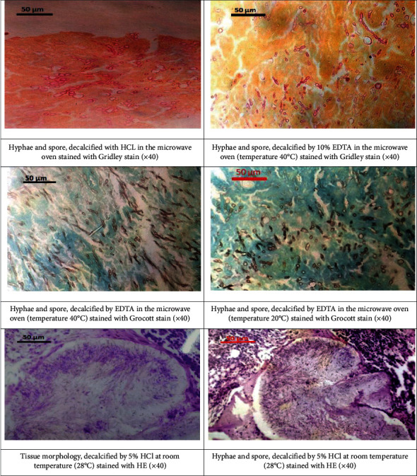
Mycetoma is a lifelong granulomatous disease of subcutaneous tissues and bones. Histopathology is a substantiated indicative method based on the assumption of a definitive diagnosis of mycetoma. It requires efficient processing of tissues including bone decalcification. The decalcification process must ensure complete removal of calcium and also a proper preservation of tissue and microorganisms' staining ability. Objectives. To compare the conventional method used in decalcification with the microwave method using different decalcification solutions. Different characteristics were tested, including the speed of decalcification and morphological and fungal preservation in bone tissue affected with mycetoma. Materials and Methods. Three decalcification solutions were employed to remove calcium from 50 bone tissue samples affected with mycetoma, including 10% neutral buffered EDTA (pH 7.4), 5% nitric acid, and 5% hydrochloric acid. Conventional and microwave methods were used. Haematoxylin-eosin (HE) stain, Gridley's stain, and Grocott hexamine-silver stain were employed to evaluate the bone and fungi morphologies. Results. The decalcification time of the conventional method compared with the microwave method with 10% EDTA (pH 7.4) took 120 hours and 29 hours, while 5% hydrochloric acid and 5% nitric acid took 8 hours and 3 hours, separately. Also, 10% EDTA is the best decalcifying agent for HE staining and fungal stains. 5% hydrochloric acid and 5% nitric acid can be used for fungal staining. Conclusion. The current study investigated the effects of different decalcifying agents as well as two decalcification procedures on the preservation of the bone structure and fungal staining, which will help to develop suitable protocols for the analyses of the bone tissue affected with mycetoma infection.


 分享
分享
 求助内容:
求助内容: 应助结果提醒方式:
 扫码关注我们
