下载PDF
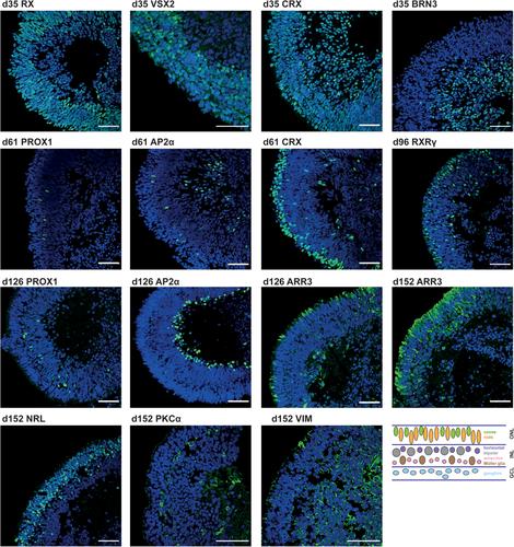
{"title":"三维视网膜类器官分化方案,免疫染色和信号定量","authors":"Hannah Döpper, Julia Menges, Morgane Bozet, Alexandra Brenzel, Dietmar Lohmann, Laura Steenpass, Deniz Kanber","doi":"10.1002/cpsc.120","DOIUrl":null,"url":null,"abstract":"<p>Structures resembling whole organs, called organoids, are generated using pluripotent stem cells and 3D culturing methods. This relies on the ability of cells to self-reorganize after dissociation. In combination with certain supplemented factors, differentiation can be directed toward the formation of several organ-like structures. Here, a protocol for the generation of retinal organoids containing all seven retinal cell types is described. This protocol does not depend on Matrigel, and by keeping the organoids single and independent at all times, fusion is prevented and monitoring of differentiation is improved. Comprehensive phenotypic characterization of the in vitro–generated retinal organoids is achieved by the protocol for immunostaining outlined here. By comparing different stages of retinal organoids, the decrease and increase of certain cell populations can be determined. In order to be able to detect even small differences, it is necessary to quantify the immunofluorescent signals, for which we have provided a detailed protocol describing signal quantitation using the image-processing program Fiji. © 2020 The Authors.</p><p><b>Basic Protocol 1</b>: Differentiation protocol for 3D retinal organoids</p><p><b>Basic Protocol 2</b>: Immunostaining protocol for cryosections of retinal organoids</p><p><b>Support Protocol</b>: Embedding and sectioning protocol for 3D retinal organoids</p><p><b>Basic Protocol 3</b>: Quantitation protocol using Fiji</p>","PeriodicalId":53703,"journal":{"name":"Current Protocols in Stem Cell Biology","volume":"55 1","pages":""},"PeriodicalIF":0.0000,"publicationDate":"2020-09-21","publicationTypes":"Journal Article","fieldsOfStudy":null,"isOpenAccess":false,"openAccessPdf":"https://sci-hub-pdf.com/10.1002/cpsc.120","citationCount":"8","resultStr":"{\"title\":\"Differentiation Protocol for 3D Retinal Organoids, Immunostaining and Signal Quantitation\",\"authors\":\"Hannah Döpper, Julia Menges, Morgane Bozet, Alexandra Brenzel, Dietmar Lohmann, Laura Steenpass, Deniz Kanber\",\"doi\":\"10.1002/cpsc.120\",\"DOIUrl\":null,\"url\":null,\"abstract\":\"<p>Structures resembling whole organs, called organoids, are generated using pluripotent stem cells and 3D culturing methods. This relies on the ability of cells to self-reorganize after dissociation. In combination with certain supplemented factors, differentiation can be directed toward the formation of several organ-like structures. Here, a protocol for the generation of retinal organoids containing all seven retinal cell types is described. This protocol does not depend on Matrigel, and by keeping the organoids single and independent at all times, fusion is prevented and monitoring of differentiation is improved. Comprehensive phenotypic characterization of the in vitro–generated retinal organoids is achieved by the protocol for immunostaining outlined here. By comparing different stages of retinal organoids, the decrease and increase of certain cell populations can be determined. In order to be able to detect even small differences, it is necessary to quantify the immunofluorescent signals, for which we have provided a detailed protocol describing signal quantitation using the image-processing program Fiji. © 2020 The Authors.</p><p><b>Basic Protocol 1</b>: Differentiation protocol for 3D retinal organoids</p><p><b>Basic Protocol 2</b>: Immunostaining protocol for cryosections of retinal organoids</p><p><b>Support Protocol</b>: Embedding and sectioning protocol for 3D retinal organoids</p><p><b>Basic Protocol 3</b>: Quantitation protocol using Fiji</p>\",\"PeriodicalId\":53703,\"journal\":{\"name\":\"Current Protocols in Stem Cell Biology\",\"volume\":\"55 1\",\"pages\":\"\"},\"PeriodicalIF\":0.0000,\"publicationDate\":\"2020-09-21\",\"publicationTypes\":\"Journal Article\",\"fieldsOfStudy\":null,\"isOpenAccess\":false,\"openAccessPdf\":\"https://sci-hub-pdf.com/10.1002/cpsc.120\",\"citationCount\":\"8\",\"resultStr\":null,\"platform\":\"Semanticscholar\",\"paperid\":null,\"PeriodicalName\":\"Current Protocols in Stem Cell Biology\",\"FirstCategoryId\":\"1085\",\"ListUrlMain\":\"https://onlinelibrary.wiley.com/doi/10.1002/cpsc.120\",\"RegionNum\":0,\"RegionCategory\":null,\"ArticlePicture\":[],\"TitleCN\":null,\"AbstractTextCN\":null,\"PMCID\":null,\"EPubDate\":\"\",\"PubModel\":\"\",\"JCR\":\"Q2\",\"JCRName\":\"Biochemistry, Genetics and Molecular Biology\",\"Score\":null,\"Total\":0}","platform":"Semanticscholar","paperid":null,"PeriodicalName":"Current Protocols in Stem Cell Biology","FirstCategoryId":"1085","ListUrlMain":"https://onlinelibrary.wiley.com/doi/10.1002/cpsc.120","RegionNum":0,"RegionCategory":null,"ArticlePicture":[],"TitleCN":null,"AbstractTextCN":null,"PMCID":null,"EPubDate":"","PubModel":"","JCR":"Q2","JCRName":"Biochemistry, Genetics and Molecular Biology","Score":null,"Total":0}
引用次数: 8
引用
批量引用


