{"title":"微计算机断层扫描病理诊断支持系统的研制。","authors":"Tomonari Hayakawa, Atsushi Teramoto, Yuka Kiriyama, Tetsuya Tsukamoto, Ayumi Yamada, Kuniaki Saito, Hiroshi Fujita","doi":"10.1267/ahc.20-00033","DOIUrl":null,"url":null,"abstract":"<p><p>In pathological diagnosis, the cutting position of pathological materials is subjectively determined by pathologists. This leads to a low cutting accuracy, which in turn may lead to incorrect diagnoses. In this study, we developed a system that supports the determination of the cutting position by visualizing and analyzing the internal structure of pathological material using micro-computed tomography (CT) before cutting. This system consists of a dedicated micro-CT and cutting support software. The micro-CT system has a fixture for fixing the target, enabling the scanning of easily deformable pathological materials. In the cutting support software, a function that interactively selects the extraction plane while displaying the volume rendering image and outputs a pseudo-histological image was implemented. Our results confirmed that the pseudo-histological image showed the fine structure inside the organ and that the latter image was highly consistent with the pathological image.</p>","PeriodicalId":6888,"journal":{"name":"Acta Histochemica Et Cytochemica","volume":"54 2","pages":"49-56"},"PeriodicalIF":1.8000,"publicationDate":"2021-04-28","publicationTypes":"Journal Article","fieldsOfStudy":null,"isOpenAccess":false,"openAccessPdf":"https://ftp.ncbi.nlm.nih.gov/pub/pmc/oa_pdf/70/4c/ahc-054-49.PMC8116619.pdf","citationCount":"1","resultStr":"{\"title\":\"Development of Pathological Diagnosis Support System Using Micro-computed Tomography.\",\"authors\":\"Tomonari Hayakawa, Atsushi Teramoto, Yuka Kiriyama, Tetsuya Tsukamoto, Ayumi Yamada, Kuniaki Saito, Hiroshi Fujita\",\"doi\":\"10.1267/ahc.20-00033\",\"DOIUrl\":null,\"url\":null,\"abstract\":\"<p><p>In pathological diagnosis, the cutting position of pathological materials is subjectively determined by pathologists. This leads to a low cutting accuracy, which in turn may lead to incorrect diagnoses. In this study, we developed a system that supports the determination of the cutting position by visualizing and analyzing the internal structure of pathological material using micro-computed tomography (CT) before cutting. This system consists of a dedicated micro-CT and cutting support software. The micro-CT system has a fixture for fixing the target, enabling the scanning of easily deformable pathological materials. In the cutting support software, a function that interactively selects the extraction plane while displaying the volume rendering image and outputs a pseudo-histological image was implemented. Our results confirmed that the pseudo-histological image showed the fine structure inside the organ and that the latter image was highly consistent with the pathological image.</p>\",\"PeriodicalId\":6888,\"journal\":{\"name\":\"Acta Histochemica Et Cytochemica\",\"volume\":\"54 2\",\"pages\":\"49-56\"},\"PeriodicalIF\":1.8000,\"publicationDate\":\"2021-04-28\",\"publicationTypes\":\"Journal Article\",\"fieldsOfStudy\":null,\"isOpenAccess\":false,\"openAccessPdf\":\"https://ftp.ncbi.nlm.nih.gov/pub/pmc/oa_pdf/70/4c/ahc-054-49.PMC8116619.pdf\",\"citationCount\":\"1\",\"resultStr\":null,\"platform\":\"Semanticscholar\",\"paperid\":null,\"PeriodicalName\":\"Acta Histochemica Et Cytochemica\",\"FirstCategoryId\":\"99\",\"ListUrlMain\":\"https://doi.org/10.1267/ahc.20-00033\",\"RegionNum\":4,\"RegionCategory\":\"生物学\",\"ArticlePicture\":[],\"TitleCN\":null,\"AbstractTextCN\":null,\"PMCID\":null,\"EPubDate\":\"2021/4/22 0:00:00\",\"PubModel\":\"Epub\",\"JCR\":\"Q4\",\"JCRName\":\"CELL BIOLOGY\",\"Score\":null,\"Total\":0}","platform":"Semanticscholar","paperid":null,"PeriodicalName":"Acta Histochemica Et Cytochemica","FirstCategoryId":"99","ListUrlMain":"https://doi.org/10.1267/ahc.20-00033","RegionNum":4,"RegionCategory":"生物学","ArticlePicture":[],"TitleCN":null,"AbstractTextCN":null,"PMCID":null,"EPubDate":"2021/4/22 0:00:00","PubModel":"Epub","JCR":"Q4","JCRName":"CELL BIOLOGY","Score":null,"Total":0}
Development of Pathological Diagnosis Support System Using Micro-computed Tomography.
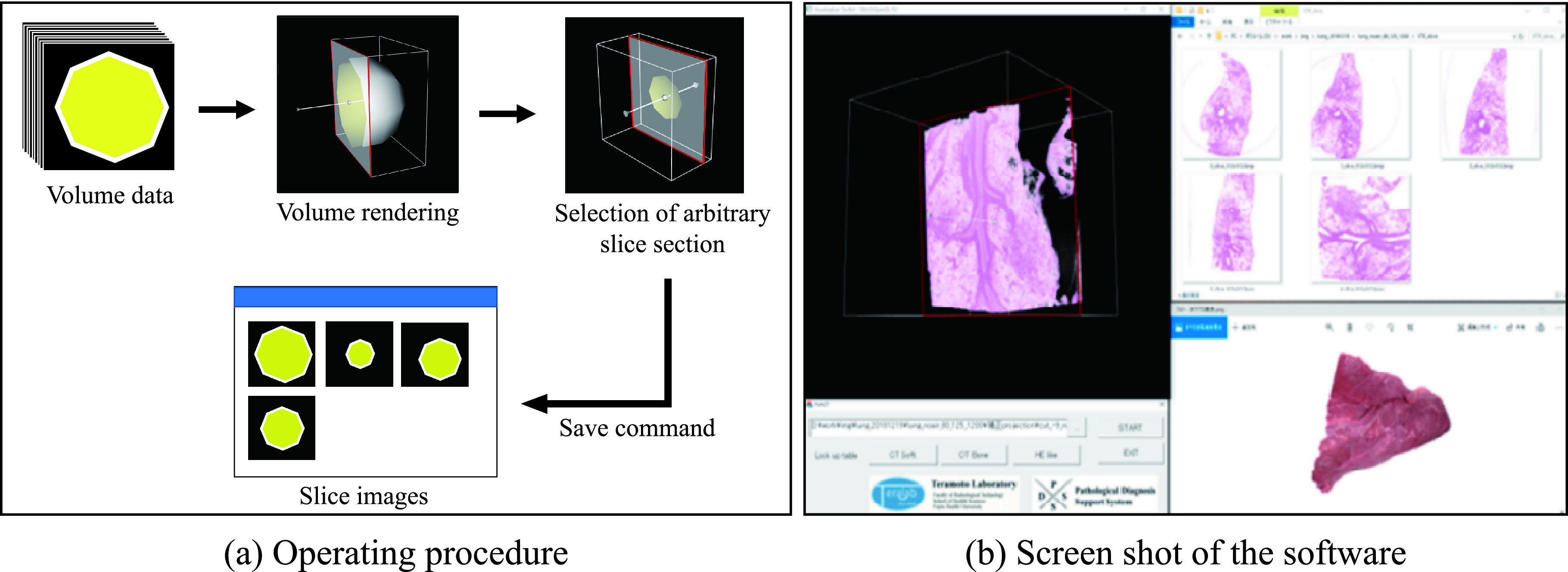
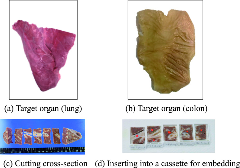
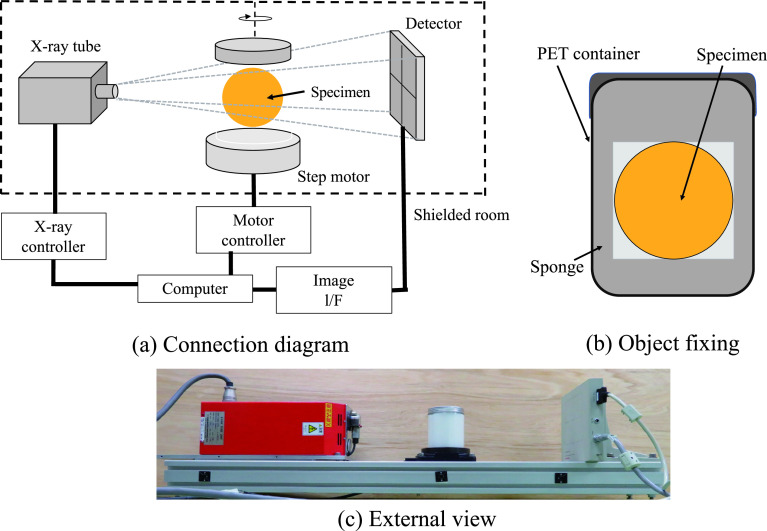
In pathological diagnosis, the cutting position of pathological materials is subjectively determined by pathologists. This leads to a low cutting accuracy, which in turn may lead to incorrect diagnoses. In this study, we developed a system that supports the determination of the cutting position by visualizing and analyzing the internal structure of pathological material using micro-computed tomography (CT) before cutting. This system consists of a dedicated micro-CT and cutting support software. The micro-CT system has a fixture for fixing the target, enabling the scanning of easily deformable pathological materials. In the cutting support software, a function that interactively selects the extraction plane while displaying the volume rendering image and outputs a pseudo-histological image was implemented. Our results confirmed that the pseudo-histological image showed the fine structure inside the organ and that the latter image was highly consistent with the pathological image.
期刊介绍:
Acta Histochemica et Cytochemica is the official online journal of the Japan Society of Histochemistry and Cytochemistry. It is intended primarily for rapid publication of concise, original articles in the fields of histochemistry and cytochemistry. Manuscripts oriented towards methodological subjects that contain significant technical advances in these fields are also welcome. Manuscripts in English are accepted from investigators in any country, whether or not they are members of the Japan Society of Histochemistry and Cytochemistry. Manuscripts should be original work that has not been previously published and is not being considered for publication elsewhere, with the exception of abstracts. Manuscripts with essentially the same content as a paper that has been published or accepted, or is under consideration for publication, will not be considered. All submitted papers will be peer-reviewed by at least two referees selected by an appropriate Associate Editor. Acceptance is based on scientific significance, originality, and clarity. When required, a revised manuscript should be submitted within 3 months, otherwise it will be considered to be a new submission. The Editor-in-Chief will make all final decisions regarding acceptance.


 分享
分享
 求助内容:
求助内容: 应助结果提醒方式:
 扫码关注我们
