Riwanti Estiasari, Adisresti Diwyacitta, Muhammad Sidik, Ni Nengah Rida Ariarini, Freddy Sitorus, Saraf Shafa Marwadhani, Kartika Maharani, Darma Imran, Reza Aditya Arpandy, David Pangeran, Manfaluthy Hakim
{"title":"评价多发性硬化症视网膜结构和视神经功能改变:1年随访的纵向研究。","authors":"Riwanti Estiasari, Adisresti Diwyacitta, Muhammad Sidik, Ni Nengah Rida Ariarini, Freddy Sitorus, Saraf Shafa Marwadhani, Kartika Maharani, Darma Imran, Reza Aditya Arpandy, David Pangeran, Manfaluthy Hakim","doi":"10.1155/2021/5573839","DOIUrl":null,"url":null,"abstract":"<p><strong>Background: </strong>Multiple sclerosis (MS) is an autoimmune disease characterized by inflammation and demyelination of the central nervous system which often involves the optic nerve even though only 20% of the patients experience optic neuritis (ON).</p><p><strong>Objective: </strong>This study aims to compare the retinal structure and optic nerve function between patients with MS and healthy controls (HCs), evaluate optic nerve alterations in MS over 1-year follow-up, and analyze its correlations with disease duration, number of relapses, degree of disability, and different subtypes.</p><p><strong>Methods: </strong>This is a prospective cohort study involving 58 eyes of MS patients. Optic nerve function was evaluated with best-corrected visual acuity (BCVA), contrast sensitivity, and P100 latency, while the retinal structure was evaluated from the GCIPL and RNFL thickness measured with optical coherence tomography (OCT) and fundus photography.</p><p><strong>Results: </strong>The MS group had lower BCVA (<i>p</i>=0.001), contrast sensitivity (<i>p</i> < 0.001), mean GCIPL thickness (<i>p</i> < 0.001), and mean RNFL thickness (<i>p</i> < 0.001) than HC. At 6 and 12 months of observations, GCIPL and RNFL (nasal quadrant) of MS patients decreased significantly (<i>p</i>=0.007 and <i>p</i>=0.004, respectively). Disease duration and the number of relapses correlated with delayed P100 latency (<i>r</i> = -0.61, <i>p</i> < 0.001 and <i>r</i> = -0.46, <i>p</i>=0.02). GCIPL and RNFL in the SPMS subtype were thinner than in RRMS.</p><p><strong>Conclusions: </strong>The retinal structure and optic nerve function of MS patients are worse than those of normal individuals. GCIPL and RNFL thinning occurs at 6 and 12 months but do not correlate with disease duration, the number of relapses, and degree of disability.</p>","PeriodicalId":19124,"journal":{"name":"Neurology Research International","volume":"2021 ","pages":"5573839"},"PeriodicalIF":2.8000,"publicationDate":"2021-06-17","publicationTypes":"Journal Article","fieldsOfStudy":null,"isOpenAccess":false,"openAccessPdf":"https://www.ncbi.nlm.nih.gov/pmc/articles/PMC8225456/pdf/","citationCount":"4","resultStr":"{\"title\":\"Evaluation of Retinal Structure and Optic Nerve Function Changes in Multiple Sclerosis: Longitudinal Study with 1-Year Follow-Up.\",\"authors\":\"Riwanti Estiasari, Adisresti Diwyacitta, Muhammad Sidik, Ni Nengah Rida Ariarini, Freddy Sitorus, Saraf Shafa Marwadhani, Kartika Maharani, Darma Imran, Reza Aditya Arpandy, David Pangeran, Manfaluthy Hakim\",\"doi\":\"10.1155/2021/5573839\",\"DOIUrl\":null,\"url\":null,\"abstract\":\"<p><strong>Background: </strong>Multiple sclerosis (MS) is an autoimmune disease characterized by inflammation and demyelination of the central nervous system which often involves the optic nerve even though only 20% of the patients experience optic neuritis (ON).</p><p><strong>Objective: </strong>This study aims to compare the retinal structure and optic nerve function between patients with MS and healthy controls (HCs), evaluate optic nerve alterations in MS over 1-year follow-up, and analyze its correlations with disease duration, number of relapses, degree of disability, and different subtypes.</p><p><strong>Methods: </strong>This is a prospective cohort study involving 58 eyes of MS patients. Optic nerve function was evaluated with best-corrected visual acuity (BCVA), contrast sensitivity, and P100 latency, while the retinal structure was evaluated from the GCIPL and RNFL thickness measured with optical coherence tomography (OCT) and fundus photography.</p><p><strong>Results: </strong>The MS group had lower BCVA (<i>p</i>=0.001), contrast sensitivity (<i>p</i> < 0.001), mean GCIPL thickness (<i>p</i> < 0.001), and mean RNFL thickness (<i>p</i> < 0.001) than HC. At 6 and 12 months of observations, GCIPL and RNFL (nasal quadrant) of MS patients decreased significantly (<i>p</i>=0.007 and <i>p</i>=0.004, respectively). Disease duration and the number of relapses correlated with delayed P100 latency (<i>r</i> = -0.61, <i>p</i> < 0.001 and <i>r</i> = -0.46, <i>p</i>=0.02). GCIPL and RNFL in the SPMS subtype were thinner than in RRMS.</p><p><strong>Conclusions: </strong>The retinal structure and optic nerve function of MS patients are worse than those of normal individuals. GCIPL and RNFL thinning occurs at 6 and 12 months but do not correlate with disease duration, the number of relapses, and degree of disability.</p>\",\"PeriodicalId\":19124,\"journal\":{\"name\":\"Neurology Research International\",\"volume\":\"2021 \",\"pages\":\"5573839\"},\"PeriodicalIF\":2.8000,\"publicationDate\":\"2021-06-17\",\"publicationTypes\":\"Journal Article\",\"fieldsOfStudy\":null,\"isOpenAccess\":false,\"openAccessPdf\":\"https://www.ncbi.nlm.nih.gov/pmc/articles/PMC8225456/pdf/\",\"citationCount\":\"4\",\"resultStr\":null,\"platform\":\"Semanticscholar\",\"paperid\":null,\"PeriodicalName\":\"Neurology Research International\",\"FirstCategoryId\":\"1085\",\"ListUrlMain\":\"https://doi.org/10.1155/2021/5573839\",\"RegionNum\":0,\"RegionCategory\":null,\"ArticlePicture\":[],\"TitleCN\":null,\"AbstractTextCN\":null,\"PMCID\":null,\"EPubDate\":\"2021/1/1 0:00:00\",\"PubModel\":\"eCollection\",\"JCR\":\"Q4\",\"JCRName\":\"NEUROSCIENCES\",\"Score\":null,\"Total\":0}","platform":"Semanticscholar","paperid":null,"PeriodicalName":"Neurology Research International","FirstCategoryId":"1085","ListUrlMain":"https://doi.org/10.1155/2021/5573839","RegionNum":0,"RegionCategory":null,"ArticlePicture":[],"TitleCN":null,"AbstractTextCN":null,"PMCID":null,"EPubDate":"2021/1/1 0:00:00","PubModel":"eCollection","JCR":"Q4","JCRName":"NEUROSCIENCES","Score":null,"Total":0}
Evaluation of Retinal Structure and Optic Nerve Function Changes in Multiple Sclerosis: Longitudinal Study with 1-Year Follow-Up.
Background: Multiple sclerosis (MS) is an autoimmune disease characterized by inflammation and demyelination of the central nervous system which often involves the optic nerve even though only 20% of the patients experience optic neuritis (ON).
Objective: This study aims to compare the retinal structure and optic nerve function between patients with MS and healthy controls (HCs), evaluate optic nerve alterations in MS over 1-year follow-up, and analyze its correlations with disease duration, number of relapses, degree of disability, and different subtypes.
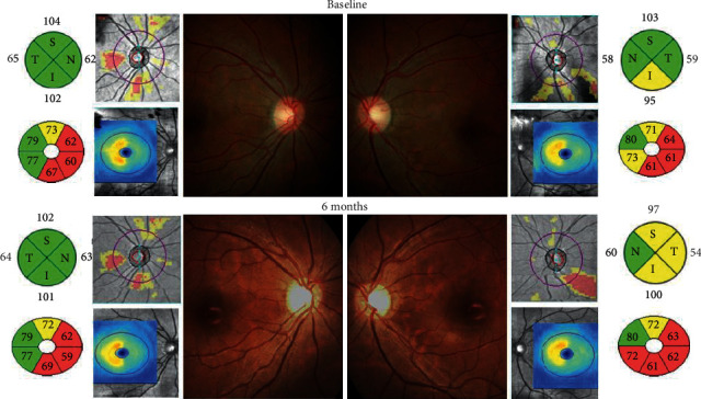
Methods: This is a prospective cohort study involving 58 eyes of MS patients. Optic nerve function was evaluated with best-corrected visual acuity (BCVA), contrast sensitivity, and P100 latency, while the retinal structure was evaluated from the GCIPL and RNFL thickness measured with optical coherence tomography (OCT) and fundus photography.
Results: The MS group had lower BCVA (p=0.001), contrast sensitivity (p < 0.001), mean GCIPL thickness (p < 0.001), and mean RNFL thickness (p < 0.001) than HC. At 6 and 12 months of observations, GCIPL and RNFL (nasal quadrant) of MS patients decreased significantly (p=0.007 and p=0.004, respectively). Disease duration and the number of relapses correlated with delayed P100 latency (r = -0.61, p < 0.001 and r = -0.46, p=0.02). GCIPL and RNFL in the SPMS subtype were thinner than in RRMS.
Conclusions: The retinal structure and optic nerve function of MS patients are worse than those of normal individuals. GCIPL and RNFL thinning occurs at 6 and 12 months but do not correlate with disease duration, the number of relapses, and degree of disability.
期刊介绍:
Neurology Research International is a peer-reviewed, Open Access journal that publishes original research articles, review articles, and clinical studies focusing on diseases of the nervous system, as well as normal neurological functioning. The journal will consider basic, translational, and clinical research, including animal models and clinical trials.


 分享
分享
 求助内容:
求助内容: 应助结果提醒方式:
 扫码关注我们
