{"title":"基于加法或减法制造的冠的边缘和内部配合。","authors":"Yasser Haddadi, Bahram Ranjkesh, Flemming Isidor, Golnosh Bahrami","doi":"10.1080/26415275.2021.1938576","DOIUrl":null,"url":null,"abstract":"<p><strong>Objective: </strong>To assess the marginal and internal fit of crowns manufactured by additive and subtractive manufacturing technique.</p><p><strong>Materials and methods: </strong>Twenty extracted teeth prepared for complete coverage crowns were scanned with an intra-oral scanner (Omnicam, DentsplySirona). For the subtractive manufacturing (SM) group, ten crowns were manufactured in a hybrid resin block (Vita Enamic, Vita Zahnfabrik). For the additive manufacturing (AM) group, the crowns were manufactured in a hybrid resin material (NextDent C&B, 3D systems). The design parameters were identical for the two groups. The marginal and internal fit (determined at the axial wall, the cusp tip and occlusally) was assessed before cementation with the replica technique and after cementation under stereomicroscope after sectioning of the crowned teeth.</p><p><strong>Results: </strong>For the SM group, the marginal fit was 91 µm (±28 µm) before cementation and 85 µm (±18 µm) after cementation. In the AM group, the marginal fit was 75 µm (±29 µm) before cementation and 71 µm (±18 µm) after cementation. The differences were not statistically significant. As regards the internal fit, the fit at the axial wall was statistically significantly better in the SM group than in the AM group (<i>p</i>=.009 before cementation and .03 after cementation). Occlusally the fit in the AM group was significantly better than in the SM group after cementation (<i>p</i><.001).</p><p><strong>Conclusion: </strong>Within the limitations of the current study, the marginal fit of additively manufactured crowns is comparable to crowns manufactured with chair-side subtractive technique and within the clinically acceptable range. As regards the internal fit no one technique was consistently superior.</p>","PeriodicalId":72378,"journal":{"name":"Biomaterial investigations in dentistry","volume":" ","pages":"87-91"},"PeriodicalIF":0.0000,"publicationDate":"2021-06-26","publicationTypes":"Journal Article","fieldsOfStudy":null,"isOpenAccess":false,"openAccessPdf":"https://sci-hub-pdf.com/10.1080/26415275.2021.1938576","citationCount":"11","resultStr":"{\"title\":\"Marginal and internal fit of crowns based on additive or subtractive manufacturing.\",\"authors\":\"Yasser Haddadi, Bahram Ranjkesh, Flemming Isidor, Golnosh Bahrami\",\"doi\":\"10.1080/26415275.2021.1938576\",\"DOIUrl\":null,\"url\":null,\"abstract\":\"<p><strong>Objective: </strong>To assess the marginal and internal fit of crowns manufactured by additive and subtractive manufacturing technique.</p><p><strong>Materials and methods: </strong>Twenty extracted teeth prepared for complete coverage crowns were scanned with an intra-oral scanner (Omnicam, DentsplySirona). For the subtractive manufacturing (SM) group, ten crowns were manufactured in a hybrid resin block (Vita Enamic, Vita Zahnfabrik). For the additive manufacturing (AM) group, the crowns were manufactured in a hybrid resin material (NextDent C&B, 3D systems). The design parameters were identical for the two groups. The marginal and internal fit (determined at the axial wall, the cusp tip and occlusally) was assessed before cementation with the replica technique and after cementation under stereomicroscope after sectioning of the crowned teeth.</p><p><strong>Results: </strong>For the SM group, the marginal fit was 91 µm (±28 µm) before cementation and 85 µm (±18 µm) after cementation. In the AM group, the marginal fit was 75 µm (±29 µm) before cementation and 71 µm (±18 µm) after cementation. The differences were not statistically significant. As regards the internal fit, the fit at the axial wall was statistically significantly better in the SM group than in the AM group (<i>p</i>=.009 before cementation and .03 after cementation). Occlusally the fit in the AM group was significantly better than in the SM group after cementation (<i>p</i><.001).</p><p><strong>Conclusion: </strong>Within the limitations of the current study, the marginal fit of additively manufactured crowns is comparable to crowns manufactured with chair-side subtractive technique and within the clinically acceptable range. As regards the internal fit no one technique was consistently superior.</p>\",\"PeriodicalId\":72378,\"journal\":{\"name\":\"Biomaterial investigations in dentistry\",\"volume\":\" \",\"pages\":\"87-91\"},\"PeriodicalIF\":0.0000,\"publicationDate\":\"2021-06-26\",\"publicationTypes\":\"Journal Article\",\"fieldsOfStudy\":null,\"isOpenAccess\":false,\"openAccessPdf\":\"https://sci-hub-pdf.com/10.1080/26415275.2021.1938576\",\"citationCount\":\"11\",\"resultStr\":null,\"platform\":\"Semanticscholar\",\"paperid\":null,\"PeriodicalName\":\"Biomaterial investigations in dentistry\",\"FirstCategoryId\":\"1085\",\"ListUrlMain\":\"https://doi.org/10.1080/26415275.2021.1938576\",\"RegionNum\":0,\"RegionCategory\":null,\"ArticlePicture\":[],\"TitleCN\":null,\"AbstractTextCN\":null,\"PMCID\":null,\"EPubDate\":\"2021/1/1 0:00:00\",\"PubModel\":\"eCollection\",\"JCR\":\"\",\"JCRName\":\"\",\"Score\":null,\"Total\":0}","platform":"Semanticscholar","paperid":null,"PeriodicalName":"Biomaterial investigations in dentistry","FirstCategoryId":"1085","ListUrlMain":"https://doi.org/10.1080/26415275.2021.1938576","RegionNum":0,"RegionCategory":null,"ArticlePicture":[],"TitleCN":null,"AbstractTextCN":null,"PMCID":null,"EPubDate":"2021/1/1 0:00:00","PubModel":"eCollection","JCR":"","JCRName":"","Score":null,"Total":0}
Marginal and internal fit of crowns based on additive or subtractive manufacturing.
Objective: To assess the marginal and internal fit of crowns manufactured by additive and subtractive manufacturing technique.
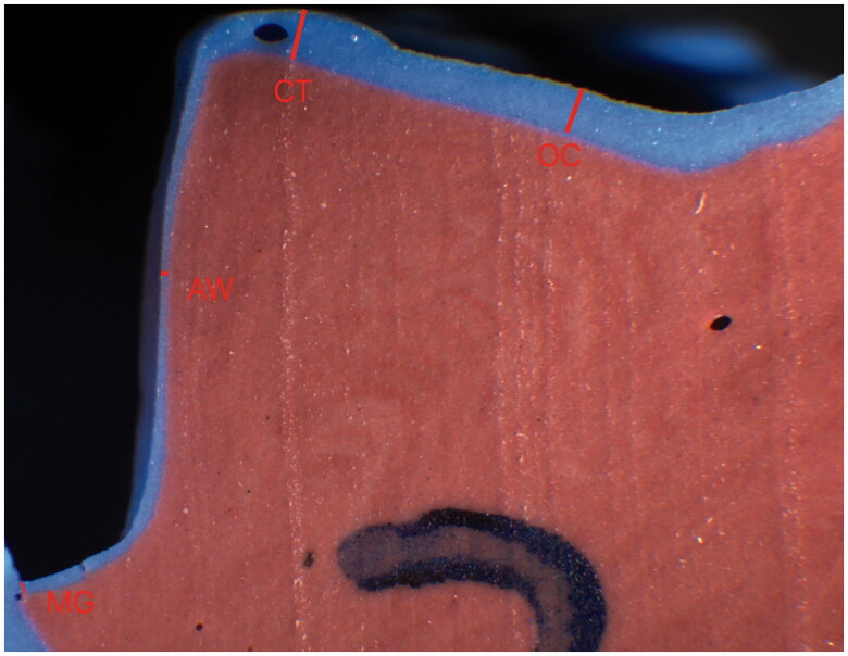
Materials and methods: Twenty extracted teeth prepared for complete coverage crowns were scanned with an intra-oral scanner (Omnicam, DentsplySirona). For the subtractive manufacturing (SM) group, ten crowns were manufactured in a hybrid resin block (Vita Enamic, Vita Zahnfabrik). For the additive manufacturing (AM) group, the crowns were manufactured in a hybrid resin material (NextDent C&B, 3D systems). The design parameters were identical for the two groups. The marginal and internal fit (determined at the axial wall, the cusp tip and occlusally) was assessed before cementation with the replica technique and after cementation under stereomicroscope after sectioning of the crowned teeth.
Results: For the SM group, the marginal fit was 91 µm (±28 µm) before cementation and 85 µm (±18 µm) after cementation. In the AM group, the marginal fit was 75 µm (±29 µm) before cementation and 71 µm (±18 µm) after cementation. The differences were not statistically significant. As regards the internal fit, the fit at the axial wall was statistically significantly better in the SM group than in the AM group (p=.009 before cementation and .03 after cementation). Occlusally the fit in the AM group was significantly better than in the SM group after cementation (p<.001).
Conclusion: Within the limitations of the current study, the marginal fit of additively manufactured crowns is comparable to crowns manufactured with chair-side subtractive technique and within the clinically acceptable range. As regards the internal fit no one technique was consistently superior.


 分享
分享

 求助内容:
求助内容: 应助结果提醒方式:
 扫码关注我们
