{"title":"(4′-氨基-5′,8′-二氟-1′- h -螺旋[哌啶-4,2′-喹唑啉]-1-基)(4-[18F]氟苯基)甲烷在诱导型一氧化氮合酶PET/MR成像中的自动合成及初步评价","authors":"Skye Hsin-Hsien Yeh, Wen-Sheng Huang, Chuang-Hsin Chiu, Chuan-Lin Chen, Hui-Ting Chen, Dae Yoon Chi, Zhengxing Ge, Tsung-Hsun Yu, Pao-Yeh Wang, Yu-Yeh Kuo, Chun-Tse Hung, Geng-Ying Li, Chi-Wei Chang","doi":"10.1155/2021/9996125","DOIUrl":null,"url":null,"abstract":"<p><strong>Background: </strong>Inducible nitric oxide synthase (iNOS) plays a crucial role in neuroinflammation, especially microglial activity, and may potentially represent a useful biomarker of neuroinflammation. In this study, we carefully defined a strategic plan to develop iNOS-targeted molecular PET imaging using (4'-amino-5',8'-difluoro-1'H-spiro[piperidine-4,2'-quinazolin]-1-yl)(4-fluorophenyl)methanone ([<sup>18</sup>F]FBAT) as a tracer in a mouse model of lipopolysaccharide- (LPS-) induced brain inflammation.</p><p><strong>Methods: </strong>An <i>in vitro</i> model, murine microglial BV2 cell line, was used to assess the uptake of [<sup>18</sup>F]FBAT in response to iNOS induction at the cellular level. <i>In vivo</i> whole-body dynamic PET/MR imaging was acquired in LPS-treated (5 mg/kg) and control mice. Standard uptake value (SUV), total volume of distribution (<i>V</i> <sub>t</sub>), and area under the curve (AUC) based on the [<sup>18</sup>F]FBAT PET signals were determined. The expression of iNOS was confirmed by immunohistochemistry (IHC) of brain tissues.</p><p><strong>Results: </strong>At the end of synthesis, the yield of [<sup>18</sup>F]FBAT was 2.2-3.1% (EOS), radiochemical purity was >99%, and molar radioactivity was 125-137 GBq/<i>μ</i>mol. <i>In vitro</i>, [<sup>18</sup>F]FBAT rapidly and progressively accumulated in murine microglial BV2 cells exposed to LPS; however, [<sup>18</sup>F]FBAT accumulation was inhibited by aminoguanidine, a selective iNOS inhibitor. <i>In vivo</i> biodistribution studies of [<sup>18</sup>F]FBAT showed a significant increase in the liver and kidney on LPS-treated mice. At 3 h postinjection of LPS, <i>in vivo</i>, the [<sup>18</sup>F]FBAT accumulation ratios at 30 min post intravenous (i.v.) radiotracer injection for the whole brain, cortex, cerebellum, and brainstem were 2.16 ± 0.18, 1.53 ± 0.25, 1.41 ± 0.21, and 1.90 ± 0.12, respectively, compared to those of mice not injected with LPS. The mean area under the curve (AUC<sub>0-30min</sub>), total volume of distribution (<i>V</i> <sub>t</sub>, mL/cm<sup>3</sup>), and <i>K</i> <sub>i</sub> (influx rate) of [<sup>18</sup>F]FBAT were 1.9 ± 0.21- and 1.4 ± 0.22-fold higher in the 3 h LPS group, respectively, than in the control group. In the pharmacokinetic two-compartment model, the whole brain <i>K</i> <sub>i</sub> of [<sup>18</sup>F]FBAT was significantly higher in mice injected with LPS compared to the control group. Aminoguanidine, selective iNOS inhibitor, pretreatment significantly reduced the AUC<sub>0-30min</sub> and <i>V</i> <sub>t</sub> values in LPS-induced mice. Quantitative analysis of immunohistochemically stained brain sections confirmed iNOS was preferentially upregulated in the cerebellum and cortex of mice injected with LPS.</p><p><strong>Conclusion: </strong>An automated robotic method was established for radiosynthesis of [<sup>18</sup>F]FBAT, and the preliminary <i>in vitro</i> and <i>in vivo</i> results demonstrated the feasibility of detecting iNOS activity/expression in LPS-treated neuroinflammation by noninvasive imaging with [<sup>18</sup>F]FBAT PET/MRI.</p>","PeriodicalId":18855,"journal":{"name":"Molecular Imaging","volume":" ","pages":"9996125"},"PeriodicalIF":2.4000,"publicationDate":"2021-07-08","publicationTypes":"Journal Article","fieldsOfStudy":null,"isOpenAccess":false,"openAccessPdf":"https://www.ncbi.nlm.nih.gov/pmc/articles/PMC8328489/pdf/","citationCount":"4","resultStr":"{\"title\":\"Automated Synthesis and Initial Evaluation of (4'-Amino-5',8'-difluoro-1'H-spiro[piperidine-4,2'-quinazolin]-1-yl)(4-[<sup>18</sup>F]fluorophenyl)methanone for PET/MR Imaging of Inducible Nitric Oxide Synthase.\",\"authors\":\"Skye Hsin-Hsien Yeh, Wen-Sheng Huang, Chuang-Hsin Chiu, Chuan-Lin Chen, Hui-Ting Chen, Dae Yoon Chi, Zhengxing Ge, Tsung-Hsun Yu, Pao-Yeh Wang, Yu-Yeh Kuo, Chun-Tse Hung, Geng-Ying Li, Chi-Wei Chang\",\"doi\":\"10.1155/2021/9996125\",\"DOIUrl\":null,\"url\":null,\"abstract\":\"<p><strong>Background: </strong>Inducible nitric oxide synthase (iNOS) plays a crucial role in neuroinflammation, especially microglial activity, and may potentially represent a useful biomarker of neuroinflammation. In this study, we carefully defined a strategic plan to develop iNOS-targeted molecular PET imaging using (4'-amino-5',8'-difluoro-1'H-spiro[piperidine-4,2'-quinazolin]-1-yl)(4-fluorophenyl)methanone ([<sup>18</sup>F]FBAT) as a tracer in a mouse model of lipopolysaccharide- (LPS-) induced brain inflammation.</p><p><strong>Methods: </strong>An <i>in vitro</i> model, murine microglial BV2 cell line, was used to assess the uptake of [<sup>18</sup>F]FBAT in response to iNOS induction at the cellular level. <i>In vivo</i> whole-body dynamic PET/MR imaging was acquired in LPS-treated (5 mg/kg) and control mice. Standard uptake value (SUV), total volume of distribution (<i>V</i> <sub>t</sub>), and area under the curve (AUC) based on the [<sup>18</sup>F]FBAT PET signals were determined. The expression of iNOS was confirmed by immunohistochemistry (IHC) of brain tissues.</p><p><strong>Results: </strong>At the end of synthesis, the yield of [<sup>18</sup>F]FBAT was 2.2-3.1% (EOS), radiochemical purity was >99%, and molar radioactivity was 125-137 GBq/<i>μ</i>mol. <i>In vitro</i>, [<sup>18</sup>F]FBAT rapidly and progressively accumulated in murine microglial BV2 cells exposed to LPS; however, [<sup>18</sup>F]FBAT accumulation was inhibited by aminoguanidine, a selective iNOS inhibitor. <i>In vivo</i> biodistribution studies of [<sup>18</sup>F]FBAT showed a significant increase in the liver and kidney on LPS-treated mice. At 3 h postinjection of LPS, <i>in vivo</i>, the [<sup>18</sup>F]FBAT accumulation ratios at 30 min post intravenous (i.v.) radiotracer injection for the whole brain, cortex, cerebellum, and brainstem were 2.16 ± 0.18, 1.53 ± 0.25, 1.41 ± 0.21, and 1.90 ± 0.12, respectively, compared to those of mice not injected with LPS. The mean area under the curve (AUC<sub>0-30min</sub>), total volume of distribution (<i>V</i> <sub>t</sub>, mL/cm<sup>3</sup>), and <i>K</i> <sub>i</sub> (influx rate) of [<sup>18</sup>F]FBAT were 1.9 ± 0.21- and 1.4 ± 0.22-fold higher in the 3 h LPS group, respectively, than in the control group. In the pharmacokinetic two-compartment model, the whole brain <i>K</i> <sub>i</sub> of [<sup>18</sup>F]FBAT was significantly higher in mice injected with LPS compared to the control group. Aminoguanidine, selective iNOS inhibitor, pretreatment significantly reduced the AUC<sub>0-30min</sub> and <i>V</i> <sub>t</sub> values in LPS-induced mice. Quantitative analysis of immunohistochemically stained brain sections confirmed iNOS was preferentially upregulated in the cerebellum and cortex of mice injected with LPS.</p><p><strong>Conclusion: </strong>An automated robotic method was established for radiosynthesis of [<sup>18</sup>F]FBAT, and the preliminary <i>in vitro</i> and <i>in vivo</i> results demonstrated the feasibility of detecting iNOS activity/expression in LPS-treated neuroinflammation by noninvasive imaging with [<sup>18</sup>F]FBAT PET/MRI.</p>\",\"PeriodicalId\":18855,\"journal\":{\"name\":\"Molecular Imaging\",\"volume\":\" \",\"pages\":\"9996125\"},\"PeriodicalIF\":2.4000,\"publicationDate\":\"2021-07-08\",\"publicationTypes\":\"Journal Article\",\"fieldsOfStudy\":null,\"isOpenAccess\":false,\"openAccessPdf\":\"https://www.ncbi.nlm.nih.gov/pmc/articles/PMC8328489/pdf/\",\"citationCount\":\"4\",\"resultStr\":null,\"platform\":\"Semanticscholar\",\"paperid\":null,\"PeriodicalName\":\"Molecular Imaging\",\"FirstCategoryId\":\"3\",\"ListUrlMain\":\"https://doi.org/10.1155/2021/9996125\",\"RegionNum\":4,\"RegionCategory\":\"医学\",\"ArticlePicture\":[],\"TitleCN\":null,\"AbstractTextCN\":null,\"PMCID\":null,\"EPubDate\":\"2021/1/1 0:00:00\",\"PubModel\":\"eCollection\",\"JCR\":\"Q3\",\"JCRName\":\"BIOCHEMICAL RESEARCH METHODS\",\"Score\":null,\"Total\":0}","platform":"Semanticscholar","paperid":null,"PeriodicalName":"Molecular Imaging","FirstCategoryId":"3","ListUrlMain":"https://doi.org/10.1155/2021/9996125","RegionNum":4,"RegionCategory":"医学","ArticlePicture":[],"TitleCN":null,"AbstractTextCN":null,"PMCID":null,"EPubDate":"2021/1/1 0:00:00","PubModel":"eCollection","JCR":"Q3","JCRName":"BIOCHEMICAL RESEARCH METHODS","Score":null,"Total":0}
引用次数: 4
摘要
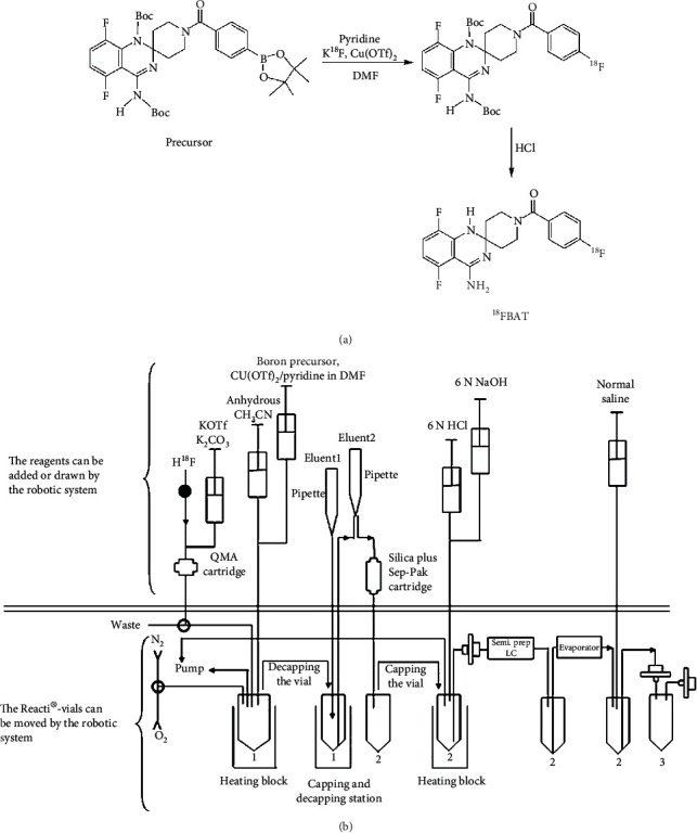
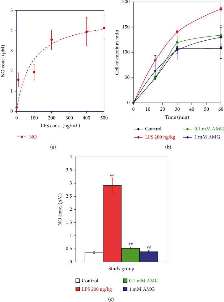
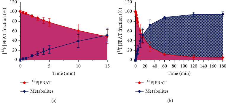
背景:诱导型一氧化氮合酶(iNOS)在神经炎症,尤其是小胶质细胞活性中起着至关重要的作用,可能是神经炎症的一个有用的生物标志物。在本研究中,我们精心制定了一项战略计划,利用(4'-氨基-5',8'-二氟-1' h -螺[哌啶-4,2'-喹唑啉]-1-基)(4-氟苯基)甲烷([18F]FBAT)作为示踪剂,在脂多糖(LPS-)诱导的脑炎症小鼠模型中开发inos靶向分子PET成像。方法:采用小鼠小胶质BV2细胞系体外模型,在细胞水平评估iNOS诱导下[18F]FBAT的摄取。在lps处理(5 mg/kg)和对照小鼠体内获得全身动态PET/MR成像。根据[18F]FBAT PET信号确定标准摄取值(SUV)、分布总量(V t)和曲线下面积(AUC)。脑组织免疫组化(IHC)证实iNOS的表达。结果:合成结束时,[18F]FBAT的产率为2.2 ~ 3.1% (EOS),放射化学纯度>99%,摩尔放射性为125 ~ 137 GBq/μmol。在体外,[18F]FBAT在暴露于LPS的小鼠小胶质BV2细胞中迅速渐进地积累;然而,[18F]选择性iNOS抑制剂氨基胍可抑制FBAT的积累。体内生物分布研究表明[18F]FBAT在lps处理小鼠的肝脏和肾脏中显著增加。注射LPS后3 h,与未注射LPS小鼠相比,静脉注射放射性示踪剂后30 min全脑、皮质、小脑和脑干的[18F]FBAT积累比分别为2.16±0.18、1.53±0.25、1.41±0.21和1.90±0.12。3 h LPS组[18F]FBAT的平均曲线下面积(AUC0-30min)、总分布容积(V t, mL/cm3)和ki(内流率)分别比对照组高1.9±0.21倍和1.4±0.22倍。在药代动力学双室模型中,注射LPS小鼠[18F]FBAT的全脑ki明显高于对照组。选择性iNOS抑制剂氨基胍预处理可显著降低lps诱导小鼠的AUC0-30min和vt值。免疫组织化学染色的脑切片定量分析证实,注射LPS小鼠的小脑和皮层iNOS优先上调。结论:建立了一种自动化机器人放射合成[18F]FBAT的方法,初步的体外和体内实验结果表明,通过[18F]FBAT PET/MRI无创成像检测lps治疗的神经炎症中iNOS活性/表达的可行性。
Automated Synthesis and Initial Evaluation of (4'-Amino-5',8'-difluoro-1'H-spiro[piperidine-4,2'-quinazolin]-1-yl)(4-[18F]fluorophenyl)methanone for PET/MR Imaging of Inducible Nitric Oxide Synthase.
Background: Inducible nitric oxide synthase (iNOS) plays a crucial role in neuroinflammation, especially microglial activity, and may potentially represent a useful biomarker of neuroinflammation. In this study, we carefully defined a strategic plan to develop iNOS-targeted molecular PET imaging using (4'-amino-5',8'-difluoro-1'H-spiro[piperidine-4,2'-quinazolin]-1-yl)(4-fluorophenyl)methanone ([18F]FBAT) as a tracer in a mouse model of lipopolysaccharide- (LPS-) induced brain inflammation.
Methods: An in vitro model, murine microglial BV2 cell line, was used to assess the uptake of [18F]FBAT in response to iNOS induction at the cellular level. In vivo whole-body dynamic PET/MR imaging was acquired in LPS-treated (5 mg/kg) and control mice. Standard uptake value (SUV), total volume of distribution (Vt), and area under the curve (AUC) based on the [18F]FBAT PET signals were determined. The expression of iNOS was confirmed by immunohistochemistry (IHC) of brain tissues.
Results: At the end of synthesis, the yield of [18F]FBAT was 2.2-3.1% (EOS), radiochemical purity was >99%, and molar radioactivity was 125-137 GBq/μmol. In vitro, [18F]FBAT rapidly and progressively accumulated in murine microglial BV2 cells exposed to LPS; however, [18F]FBAT accumulation was inhibited by aminoguanidine, a selective iNOS inhibitor. In vivo biodistribution studies of [18F]FBAT showed a significant increase in the liver and kidney on LPS-treated mice. At 3 h postinjection of LPS, in vivo, the [18F]FBAT accumulation ratios at 30 min post intravenous (i.v.) radiotracer injection for the whole brain, cortex, cerebellum, and brainstem were 2.16 ± 0.18, 1.53 ± 0.25, 1.41 ± 0.21, and 1.90 ± 0.12, respectively, compared to those of mice not injected with LPS. The mean area under the curve (AUC0-30min), total volume of distribution (Vt, mL/cm3), and Ki (influx rate) of [18F]FBAT were 1.9 ± 0.21- and 1.4 ± 0.22-fold higher in the 3 h LPS group, respectively, than in the control group. In the pharmacokinetic two-compartment model, the whole brain Ki of [18F]FBAT was significantly higher in mice injected with LPS compared to the control group. Aminoguanidine, selective iNOS inhibitor, pretreatment significantly reduced the AUC0-30min and Vt values in LPS-induced mice. Quantitative analysis of immunohistochemically stained brain sections confirmed iNOS was preferentially upregulated in the cerebellum and cortex of mice injected with LPS.
Conclusion: An automated robotic method was established for radiosynthesis of [18F]FBAT, and the preliminary in vitro and in vivo results demonstrated the feasibility of detecting iNOS activity/expression in LPS-treated neuroinflammation by noninvasive imaging with [18F]FBAT PET/MRI.
Molecular ImagingBiochemistry, Genetics and Molecular Biology-Biotechnology
自引率
3.60%
发文量
21
期刊介绍:
Molecular Imaging is a peer-reviewed, open access journal highlighting the breadth of molecular imaging research from basic science to preclinical studies to human applications. This serves both the scientific and clinical communities by disseminating novel results and concepts relevant to the biological study of normal and disease processes in both basic and translational studies ranging from mice to humans.


 分享
分享
 求助内容:
求助内容: 应助结果提醒方式:
 扫码关注我们
