Vincent A Stadelmann, Gabrielle Boyd, Martin Guillot, Jean-Guy Bienvenu, Charles Glaus, Aurore Varela
{"title":"在离体小鼠主动脉增强微ct扫描中动脉粥样硬化的自动定量。","authors":"Vincent A Stadelmann, Gabrielle Boyd, Martin Guillot, Jean-Guy Bienvenu, Charles Glaus, Aurore Varela","doi":"10.1155/2021/4998786","DOIUrl":null,"url":null,"abstract":"<p><strong>Objective: </strong>While microCT evaluation of atherosclerotic lesions in mice has been formally validated, existing image processing methods remain undisclosed. We aimed to develop and validate a reproducible image processing workflow based on phosphotungstic acid-enhanced microCT scans for the volumetric quantification of atherosclerotic lesions in entire mouse aortas. <i>Approach and Results</i>. 42 WT and 42 apolipoprotein E knockout mouse aortas were scanned. The walls, lumen, and plaque objects were segmented using dual-threshold algorithms. Aortic and plaque volumes were computed by voxel counting and lesion surface by triangulation. The results were validated against manual and histological evaluations. Knockout mice had a significant increase in plaque volume compared to wild types with a plaque to aorta volume ratio of 0.3%, 2.8%, and 9.8% at weeks 13, 18, and 26, respectively. Automatic segmentation correlated with manual (<i>r</i> <sup>2</sup> ≥ 0.89; <i>p</i> < .001) and histological evaluations (<i>r</i> <sup>2</sup> > 0.96; <i>p</i> < .001).</p><p><strong>Conclusions: </strong>The semiautomatic workflow enabled rapid quantification of atherosclerotic plaques in mice with minimal manual work.</p>","PeriodicalId":47063,"journal":{"name":"International Journal of Biomedical Imaging","volume":" ","pages":"4998786"},"PeriodicalIF":1.3000,"publicationDate":"2021-09-20","publicationTypes":"Journal Article","fieldsOfStudy":null,"isOpenAccess":false,"openAccessPdf":"https://www.ncbi.nlm.nih.gov/pmc/articles/PMC8478544/pdf/","citationCount":"3","resultStr":"{\"title\":\"Automatic Quantification of Atherosclerosis in Contrast-Enhanced MicroCT Scans of Mouse Aortas Ex Vivo.\",\"authors\":\"Vincent A Stadelmann, Gabrielle Boyd, Martin Guillot, Jean-Guy Bienvenu, Charles Glaus, Aurore Varela\",\"doi\":\"10.1155/2021/4998786\",\"DOIUrl\":null,\"url\":null,\"abstract\":\"<p><strong>Objective: </strong>While microCT evaluation of atherosclerotic lesions in mice has been formally validated, existing image processing methods remain undisclosed. We aimed to develop and validate a reproducible image processing workflow based on phosphotungstic acid-enhanced microCT scans for the volumetric quantification of atherosclerotic lesions in entire mouse aortas. <i>Approach and Results</i>. 42 WT and 42 apolipoprotein E knockout mouse aortas were scanned. The walls, lumen, and plaque objects were segmented using dual-threshold algorithms. Aortic and plaque volumes were computed by voxel counting and lesion surface by triangulation. The results were validated against manual and histological evaluations. Knockout mice had a significant increase in plaque volume compared to wild types with a plaque to aorta volume ratio of 0.3%, 2.8%, and 9.8% at weeks 13, 18, and 26, respectively. Automatic segmentation correlated with manual (<i>r</i> <sup>2</sup> ≥ 0.89; <i>p</i> < .001) and histological evaluations (<i>r</i> <sup>2</sup> > 0.96; <i>p</i> < .001).</p><p><strong>Conclusions: </strong>The semiautomatic workflow enabled rapid quantification of atherosclerotic plaques in mice with minimal manual work.</p>\",\"PeriodicalId\":47063,\"journal\":{\"name\":\"International Journal of Biomedical Imaging\",\"volume\":\" \",\"pages\":\"4998786\"},\"PeriodicalIF\":1.3000,\"publicationDate\":\"2021-09-20\",\"publicationTypes\":\"Journal Article\",\"fieldsOfStudy\":null,\"isOpenAccess\":false,\"openAccessPdf\":\"https://www.ncbi.nlm.nih.gov/pmc/articles/PMC8478544/pdf/\",\"citationCount\":\"3\",\"resultStr\":null,\"platform\":\"Semanticscholar\",\"paperid\":null,\"PeriodicalName\":\"International Journal of Biomedical Imaging\",\"FirstCategoryId\":\"1085\",\"ListUrlMain\":\"https://doi.org/10.1155/2021/4998786\",\"RegionNum\":0,\"RegionCategory\":null,\"ArticlePicture\":[],\"TitleCN\":null,\"AbstractTextCN\":null,\"PMCID\":null,\"EPubDate\":\"2021/1/1 0:00:00\",\"PubModel\":\"eCollection\",\"JCR\":\"Q2\",\"JCRName\":\"ENGINEERING, BIOMEDICAL\",\"Score\":null,\"Total\":0}","platform":"Semanticscholar","paperid":null,"PeriodicalName":"International Journal of Biomedical Imaging","FirstCategoryId":"1085","ListUrlMain":"https://doi.org/10.1155/2021/4998786","RegionNum":0,"RegionCategory":null,"ArticlePicture":[],"TitleCN":null,"AbstractTextCN":null,"PMCID":null,"EPubDate":"2021/1/1 0:00:00","PubModel":"eCollection","JCR":"Q2","JCRName":"ENGINEERING, BIOMEDICAL","Score":null,"Total":0}
Automatic Quantification of Atherosclerosis in Contrast-Enhanced MicroCT Scans of Mouse Aortas Ex Vivo.
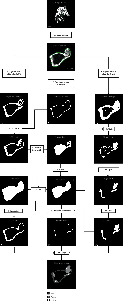
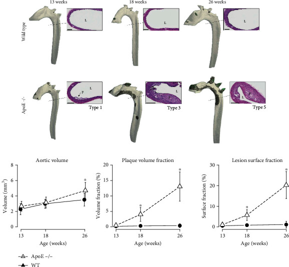
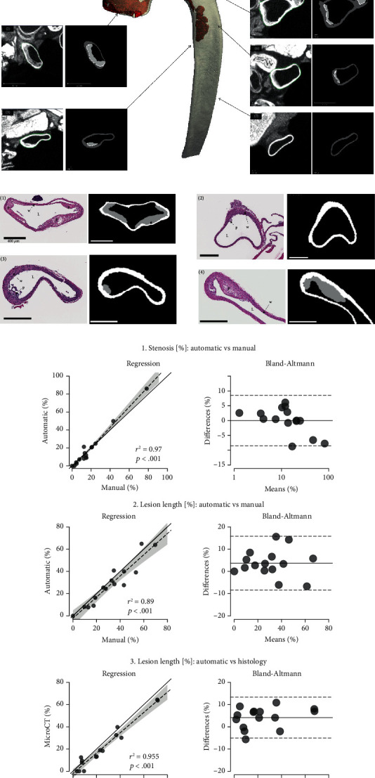
Objective: While microCT evaluation of atherosclerotic lesions in mice has been formally validated, existing image processing methods remain undisclosed. We aimed to develop and validate a reproducible image processing workflow based on phosphotungstic acid-enhanced microCT scans for the volumetric quantification of atherosclerotic lesions in entire mouse aortas. Approach and Results. 42 WT and 42 apolipoprotein E knockout mouse aortas were scanned. The walls, lumen, and plaque objects were segmented using dual-threshold algorithms. Aortic and plaque volumes were computed by voxel counting and lesion surface by triangulation. The results were validated against manual and histological evaluations. Knockout mice had a significant increase in plaque volume compared to wild types with a plaque to aorta volume ratio of 0.3%, 2.8%, and 9.8% at weeks 13, 18, and 26, respectively. Automatic segmentation correlated with manual (r2 ≥ 0.89; p < .001) and histological evaluations (r2 > 0.96; p < .001).
Conclusions: The semiautomatic workflow enabled rapid quantification of atherosclerotic plaques in mice with minimal manual work.
期刊介绍:
The International Journal of Biomedical Imaging is managed by a board of editors comprising internationally renowned active researchers. The journal is freely accessible online and also offered for purchase in print format. It employs a web-based review system to ensure swift turnaround times while maintaining high standards. In addition to regular issues, special issues are organized by guest editors. The subject areas covered include (but are not limited to):
Digital radiography and tomosynthesis
X-ray computed tomography (CT)
Magnetic resonance imaging (MRI)
Single photon emission computed tomography (SPECT)
Positron emission tomography (PET)
Ultrasound imaging
Diffuse optical tomography, coherence, fluorescence, bioluminescence tomography, impedance tomography
Neutron imaging for biomedical applications
Magnetic and optical spectroscopy, and optical biopsy
Optical, electron, scanning tunneling/atomic force microscopy
Small animal imaging
Functional, cellular, and molecular imaging
Imaging assays for screening and molecular analysis
Microarray image analysis and bioinformatics
Emerging biomedical imaging techniques
Imaging modality fusion
Biomedical imaging instrumentation
Biomedical image processing, pattern recognition, and analysis
Biomedical image visualization, compression, transmission, and storage
Imaging and modeling related to systems biology and systems biomedicine
Applied mathematics, applied physics, and chemistry related to biomedical imaging
Grid-enabling technology for biomedical imaging and informatics


 分享
分享
 求助内容:
求助内容: 应助结果提醒方式:
 扫码关注我们
