{"title":"我的耳朵在哪里?-颈椎软骨皮鳃裂残余。","authors":"Smile Kajal, Anam Ahmed, Anurag Gupta","doi":"10.4274/tao.2021.2021-6-13","DOIUrl":null,"url":null,"abstract":"A 6-year-old boy presented with the absence of pinna on the left side since birth. A cutaneous appendage emerging from the neck on the left side was discovered during the examination (Figure 1). Deep palpation revealed a firm texture like cartilage. On the left side, a primitive firm cartilage was palpated in the helix region (Figure 2). The rest of the cartilaginous framework and external auditory canal (EAC) opening was absent on the left side. On the right side, the pinna was smaller in size and loped. High resolution computed tomography (HRCT) of temporal bone showed an atretic and stenosed EAC on the left and ride side, respectively. Ossicular lump was noted on both sides on HRCT, but all inner ear structures appeared normal in both ears. Pure tone audiometry revealed moderate and mild conductive hearing loss on the left and right sides, respectively. Magnetic resonance imaging of the neck was done to rule out any branchial abnormality. It did not reveal any cyst/sinus/fistula. A provisional diagnosis of cervical chondrocutaneous branchial remnant (CCBR) with leftsided microtia and EAC atresia was made on clinico-radiological basis. After a thorough discussion with the father, the patient was planned for the excision of the cervical mass and staged pinnaplasty and canaloplasty on the left side at a later stage. Histopathological examination after excision showed elastic cartilage rests covered by normal skin consisting of epidermis, dermis, adnexal structures, and subcutaneous fat compatible with CCBR.","PeriodicalId":44240,"journal":{"name":"Turkish Archives of Otorhinolaryngology","volume":"59 3","pages":"242-243"},"PeriodicalIF":0.6000,"publicationDate":"2021-09-01","publicationTypes":"Journal Article","fieldsOfStudy":null,"isOpenAccess":false,"openAccessPdf":"https://ftp.ncbi.nlm.nih.gov/pub/pmc/oa_pdf/94/19/tao-59-242.PMC8527535.pdf","citationCount":"2","resultStr":"{\"title\":\"Where is my ear? - Cervical Chondrocutaneous Branchial Remnant.\",\"authors\":\"Smile Kajal, Anam Ahmed, Anurag Gupta\",\"doi\":\"10.4274/tao.2021.2021-6-13\",\"DOIUrl\":null,\"url\":null,\"abstract\":\"A 6-year-old boy presented with the absence of pinna on the left side since birth. A cutaneous appendage emerging from the neck on the left side was discovered during the examination (Figure 1). Deep palpation revealed a firm texture like cartilage. On the left side, a primitive firm cartilage was palpated in the helix region (Figure 2). The rest of the cartilaginous framework and external auditory canal (EAC) opening was absent on the left side. On the right side, the pinna was smaller in size and loped. High resolution computed tomography (HRCT) of temporal bone showed an atretic and stenosed EAC on the left and ride side, respectively. Ossicular lump was noted on both sides on HRCT, but all inner ear structures appeared normal in both ears. Pure tone audiometry revealed moderate and mild conductive hearing loss on the left and right sides, respectively. Magnetic resonance imaging of the neck was done to rule out any branchial abnormality. It did not reveal any cyst/sinus/fistula. A provisional diagnosis of cervical chondrocutaneous branchial remnant (CCBR) with leftsided microtia and EAC atresia was made on clinico-radiological basis. After a thorough discussion with the father, the patient was planned for the excision of the cervical mass and staged pinnaplasty and canaloplasty on the left side at a later stage. Histopathological examination after excision showed elastic cartilage rests covered by normal skin consisting of epidermis, dermis, adnexal structures, and subcutaneous fat compatible with CCBR.\",\"PeriodicalId\":44240,\"journal\":{\"name\":\"Turkish Archives of Otorhinolaryngology\",\"volume\":\"59 3\",\"pages\":\"242-243\"},\"PeriodicalIF\":0.6000,\"publicationDate\":\"2021-09-01\",\"publicationTypes\":\"Journal Article\",\"fieldsOfStudy\":null,\"isOpenAccess\":false,\"openAccessPdf\":\"https://ftp.ncbi.nlm.nih.gov/pub/pmc/oa_pdf/94/19/tao-59-242.PMC8527535.pdf\",\"citationCount\":\"2\",\"resultStr\":null,\"platform\":\"Semanticscholar\",\"paperid\":null,\"PeriodicalName\":\"Turkish Archives of Otorhinolaryngology\",\"FirstCategoryId\":\"1085\",\"ListUrlMain\":\"https://doi.org/10.4274/tao.2021.2021-6-13\",\"RegionNum\":0,\"RegionCategory\":null,\"ArticlePicture\":[],\"TitleCN\":null,\"AbstractTextCN\":null,\"PMCID\":null,\"EPubDate\":\"2021/10/15 0:00:00\",\"PubModel\":\"Epub\",\"JCR\":\"Q4\",\"JCRName\":\"OTORHINOLARYNGOLOGY\",\"Score\":null,\"Total\":0}","platform":"Semanticscholar","paperid":null,"PeriodicalName":"Turkish Archives of Otorhinolaryngology","FirstCategoryId":"1085","ListUrlMain":"https://doi.org/10.4274/tao.2021.2021-6-13","RegionNum":0,"RegionCategory":null,"ArticlePicture":[],"TitleCN":null,"AbstractTextCN":null,"PMCID":null,"EPubDate":"2021/10/15 0:00:00","PubModel":"Epub","JCR":"Q4","JCRName":"OTORHINOLARYNGOLOGY","Score":null,"Total":0}
Where is my ear? - Cervical Chondrocutaneous Branchial Remnant.
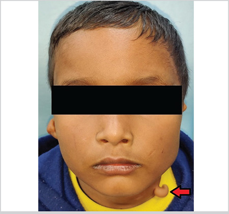
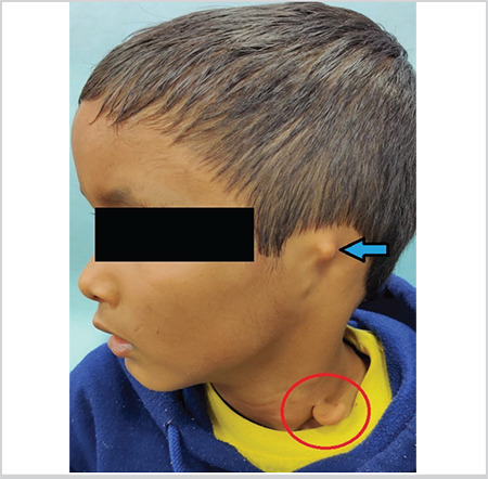
A 6-year-old boy presented with the absence of pinna on the left side since birth. A cutaneous appendage emerging from the neck on the left side was discovered during the examination (Figure 1). Deep palpation revealed a firm texture like cartilage. On the left side, a primitive firm cartilage was palpated in the helix region (Figure 2). The rest of the cartilaginous framework and external auditory canal (EAC) opening was absent on the left side. On the right side, the pinna was smaller in size and loped. High resolution computed tomography (HRCT) of temporal bone showed an atretic and stenosed EAC on the left and ride side, respectively. Ossicular lump was noted on both sides on HRCT, but all inner ear structures appeared normal in both ears. Pure tone audiometry revealed moderate and mild conductive hearing loss on the left and right sides, respectively. Magnetic resonance imaging of the neck was done to rule out any branchial abnormality. It did not reveal any cyst/sinus/fistula. A provisional diagnosis of cervical chondrocutaneous branchial remnant (CCBR) with leftsided microtia and EAC atresia was made on clinico-radiological basis. After a thorough discussion with the father, the patient was planned for the excision of the cervical mass and staged pinnaplasty and canaloplasty on the left side at a later stage. Histopathological examination after excision showed elastic cartilage rests covered by normal skin consisting of epidermis, dermis, adnexal structures, and subcutaneous fat compatible with CCBR.


 分享
分享
 求助内容:
求助内容: 应助结果提醒方式:
 扫码关注我们
