Kathryn E Keenan, Ben P Berman, Slávka Rýger, Stephen E Russek, Wen-Tung Wang, John A Butman, Dzung L Pham, Joseph Dagher
{"title":"使用旋转管模型的相位估计方法与定量易感性绘图的比较","authors":"Kathryn E Keenan, Ben P Berman, Slávka Rýger, Stephen E Russek, Wen-Tung Wang, John A Butman, Dzung L Pham, Joseph Dagher","doi":"10.1155/2021/1898461","DOIUrl":null,"url":null,"abstract":"<p><p>Quantitative Susceptibility Mapping (QSM) is an MRI tool with the potential to reveal pathological changes from magnetic susceptibility measurements. Before phase data can be used to recover susceptibility (Δ<i>χ</i>), the QSM process begins with two steps: data acquisition and phase estimation. We assess the performance of these steps, when applied without user intervention, on several variations of a phantom imaging task. We used a rotating-tube phantom with five tubes ranging from Δ<i>χ</i>=0.05 ppm to Δ<i>χ</i>=0.336 ppm. MRI data was acquired at nine angles of rotation for four different pulse sequences. The images were processed by 10 phase estimation algorithms including Laplacian, region-growing, branch-cut, temporal unwrapping, and maximum-likelihood methods, resulting in approximately 90 different combinations of data acquisition and phase estimation methods. We analyzed errors between measured and expected phases using the probability mass function and Cumulative Distribution Function. Repeatable acquisition and estimation methods were identified based on the probability of relative phase errors. For single-echo GRE and segmented EPI sequences, a region-growing method was most reliable with Pr (relative error <0.1) = 0.95 and 0.90, respectively. For multiecho sequences, a maximum-likelihood method was most reliable with Pr (relative error <0.1) = 0.97. The most repeatable multiecho methods outperformed the most repeatable single-echo methods. We found a wide range of repeatability and reproducibility for off-the-shelf MRI acquisition and phase estimation approaches, and this variability may prevent the techniques from being widely integrated in clinical workflows. The error was dominated in many cases by spatially discontinuous phase unwrapping errors. Any postprocessing applied on erroneous phase estimates, such as QSM's background field removal and dipole inversion, would suffer from error propagation. Our paradigm identifies methods that yield consistent and accurate phase estimates that would ultimately yield consistent and accurate Δ<i>χ</i> estimates.</p>","PeriodicalId":51864,"journal":{"name":"Radiology Research and Practice","volume":"2021 ","pages":"1898461"},"PeriodicalIF":1.5000,"publicationDate":"2021-11-24","publicationTypes":"Journal Article","fieldsOfStudy":null,"isOpenAccess":false,"openAccessPdf":"https://www.ncbi.nlm.nih.gov/pmc/articles/PMC8635951/pdf/","citationCount":"0","resultStr":"{\"title\":\"Comparison of Phase Estimation Methods for Quantitative Susceptibility Mapping Using a Rotating-Tube Phantom.\",\"authors\":\"Kathryn E Keenan, Ben P Berman, Slávka Rýger, Stephen E Russek, Wen-Tung Wang, John A Butman, Dzung L Pham, Joseph Dagher\",\"doi\":\"10.1155/2021/1898461\",\"DOIUrl\":null,\"url\":null,\"abstract\":\"<p><p>Quantitative Susceptibility Mapping (QSM) is an MRI tool with the potential to reveal pathological changes from magnetic susceptibility measurements. Before phase data can be used to recover susceptibility (Δ<i>χ</i>), the QSM process begins with two steps: data acquisition and phase estimation. We assess the performance of these steps, when applied without user intervention, on several variations of a phantom imaging task. We used a rotating-tube phantom with five tubes ranging from Δ<i>χ</i>=0.05 ppm to Δ<i>χ</i>=0.336 ppm. MRI data was acquired at nine angles of rotation for four different pulse sequences. The images were processed by 10 phase estimation algorithms including Laplacian, region-growing, branch-cut, temporal unwrapping, and maximum-likelihood methods, resulting in approximately 90 different combinations of data acquisition and phase estimation methods. We analyzed errors between measured and expected phases using the probability mass function and Cumulative Distribution Function. Repeatable acquisition and estimation methods were identified based on the probability of relative phase errors. For single-echo GRE and segmented EPI sequences, a region-growing method was most reliable with Pr (relative error <0.1) = 0.95 and 0.90, respectively. For multiecho sequences, a maximum-likelihood method was most reliable with Pr (relative error <0.1) = 0.97. The most repeatable multiecho methods outperformed the most repeatable single-echo methods. We found a wide range of repeatability and reproducibility for off-the-shelf MRI acquisition and phase estimation approaches, and this variability may prevent the techniques from being widely integrated in clinical workflows. The error was dominated in many cases by spatially discontinuous phase unwrapping errors. Any postprocessing applied on erroneous phase estimates, such as QSM's background field removal and dipole inversion, would suffer from error propagation. Our paradigm identifies methods that yield consistent and accurate phase estimates that would ultimately yield consistent and accurate Δ<i>χ</i> estimates.</p>\",\"PeriodicalId\":51864,\"journal\":{\"name\":\"Radiology Research and Practice\",\"volume\":\"2021 \",\"pages\":\"1898461\"},\"PeriodicalIF\":1.5000,\"publicationDate\":\"2021-11-24\",\"publicationTypes\":\"Journal Article\",\"fieldsOfStudy\":null,\"isOpenAccess\":false,\"openAccessPdf\":\"https://www.ncbi.nlm.nih.gov/pmc/articles/PMC8635951/pdf/\",\"citationCount\":\"0\",\"resultStr\":null,\"platform\":\"Semanticscholar\",\"paperid\":null,\"PeriodicalName\":\"Radiology Research and Practice\",\"FirstCategoryId\":\"1085\",\"ListUrlMain\":\"https://doi.org/10.1155/2021/1898461\",\"RegionNum\":0,\"RegionCategory\":null,\"ArticlePicture\":[],\"TitleCN\":null,\"AbstractTextCN\":null,\"PMCID\":null,\"EPubDate\":\"2021/1/1 0:00:00\",\"PubModel\":\"eCollection\",\"JCR\":\"Q2\",\"JCRName\":\"RADIOLOGY, NUCLEAR MEDICINE & MEDICAL IMAGING\",\"Score\":null,\"Total\":0}","platform":"Semanticscholar","paperid":null,"PeriodicalName":"Radiology Research and Practice","FirstCategoryId":"1085","ListUrlMain":"https://doi.org/10.1155/2021/1898461","RegionNum":0,"RegionCategory":null,"ArticlePicture":[],"TitleCN":null,"AbstractTextCN":null,"PMCID":null,"EPubDate":"2021/1/1 0:00:00","PubModel":"eCollection","JCR":"Q2","JCRName":"RADIOLOGY, NUCLEAR MEDICINE & MEDICAL IMAGING","Score":null,"Total":0}
Comparison of Phase Estimation Methods for Quantitative Susceptibility Mapping Using a Rotating-Tube Phantom.
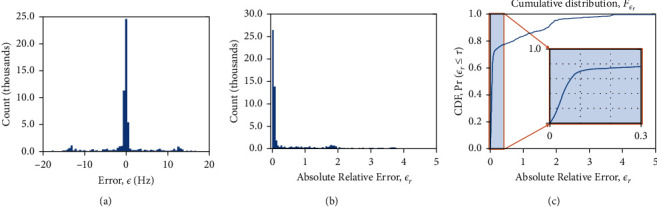
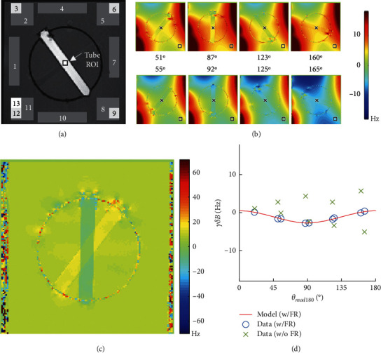
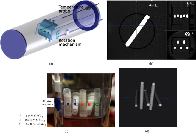
Quantitative Susceptibility Mapping (QSM) is an MRI tool with the potential to reveal pathological changes from magnetic susceptibility measurements. Before phase data can be used to recover susceptibility (Δχ), the QSM process begins with two steps: data acquisition and phase estimation. We assess the performance of these steps, when applied without user intervention, on several variations of a phantom imaging task. We used a rotating-tube phantom with five tubes ranging from Δχ=0.05 ppm to Δχ=0.336 ppm. MRI data was acquired at nine angles of rotation for four different pulse sequences. The images were processed by 10 phase estimation algorithms including Laplacian, region-growing, branch-cut, temporal unwrapping, and maximum-likelihood methods, resulting in approximately 90 different combinations of data acquisition and phase estimation methods. We analyzed errors between measured and expected phases using the probability mass function and Cumulative Distribution Function. Repeatable acquisition and estimation methods were identified based on the probability of relative phase errors. For single-echo GRE and segmented EPI sequences, a region-growing method was most reliable with Pr (relative error <0.1) = 0.95 and 0.90, respectively. For multiecho sequences, a maximum-likelihood method was most reliable with Pr (relative error <0.1) = 0.97. The most repeatable multiecho methods outperformed the most repeatable single-echo methods. We found a wide range of repeatability and reproducibility for off-the-shelf MRI acquisition and phase estimation approaches, and this variability may prevent the techniques from being widely integrated in clinical workflows. The error was dominated in many cases by spatially discontinuous phase unwrapping errors. Any postprocessing applied on erroneous phase estimates, such as QSM's background field removal and dipole inversion, would suffer from error propagation. Our paradigm identifies methods that yield consistent and accurate phase estimates that would ultimately yield consistent and accurate Δχ estimates.
期刊介绍:
Radiology Research and Practice is a peer-reviewed, Open Access journal that publishes articles on all areas of medical imaging. The journal promotes evidence-based radiology practice though the publication of original research, reviews, and clinical studies for a multidisciplinary audience. Radiology Research and Practice is archived in Portico, which provides permanent archiving for electronic scholarly journals, as well as via the LOCKSS initiative. It operates a fully open access publishing model which allows open global access to its published content. This model is supported through Article Processing Charges. For more information on Article Processing charges in gen


 分享
分享
 求助内容:
求助内容: 应助结果提醒方式:
 扫码关注我们
