Nathaly Enciso, José Amiel, Fredy Fabián-Domínguez, Jhon Pando, Nancy Rojas, Carlos Cisneros-Huamaní, Ernesto Nava, Javier Enciso
{"title":"硫乙酰胺诱导大鼠肝纤维化再生治疗模型的建立。","authors":"Nathaly Enciso, José Amiel, Fredy Fabián-Domínguez, Jhon Pando, Nancy Rojas, Carlos Cisneros-Huamaní, Ernesto Nava, Javier Enciso","doi":"10.1155/2022/2841894","DOIUrl":null,"url":null,"abstract":"<p><p>Hepatic fibrosis is caused by chronic injury due to toxic, infectious, or metabolic causes, and it may progress to cirrhosis and hepatocellular carcinoma. There is currently no antifibrotic therapy authorized for human use; however, there are promising studies using cell therapies. There are also no animal models that exactly reproduce human liver fibrosis that can be used to better understand the mechanisms of its regression and identify new targets for treatment and therapeutic approaches. On the other hand, mesenchymal stem cells (MSC) have experimentally demonstrated fibrosis regression effects, but it is necessary to have an animal model of advanced liver fibrosis to evaluate the effect of these cells. The aim of this work was to establish a protocol for the induction of advanced liver fibrosis in rats using thioacetamide (TAA), which will allow us to perform trials using MSC as a possible therapy for fibrosis regression. For this purpose, we selected 24 female rats and grouped them into three experimental groups: the control group (G-I) without treatment and groups II (G-II) and III (G-III) that received TAA by intraperitoneal injection for 24 weeks. Then, 1 × 10⁶/kg adipose mesenchymal stem cells (ASCs) were infused intravenously. Groups G-I and G-II were sacrificed 7 days after the last dose of ASC, and G-III was sacrificed 8 weeks after the last ASC infusion, all with xylazine/ketamine (40 mg/kg). The protocol used in this work established a model of advanced hepatic fibrosis as corroborated by METAVIR tests of the histological lesions; by the high levels of the markers <i>α</i>-SMA, CD68, and collagen type I; by functional alterations due to elevated markers of the hepatic lesions; and by alterations of the leukocytes, lymphocytes, and platelets. Finally, transplanted cells in the fibrous liver were detected. We conclude that TAA applied using the protocol introduced in this study induces a good model of advanced liver fibrosis in rats.</p>","PeriodicalId":313227,"journal":{"name":"Analytical Cellular Pathology (Amsterdam)","volume":" ","pages":"2841894"},"PeriodicalIF":0.0000,"publicationDate":"2022-11-12","publicationTypes":"Journal Article","fieldsOfStudy":null,"isOpenAccess":false,"openAccessPdf":"https://www.ncbi.nlm.nih.gov/pmc/articles/PMC9675604/pdf/","citationCount":"2","resultStr":"{\"title\":\"Model of Liver Fibrosis Induction by Thioacetamide in Rats for Regenerative Therapy Studies.\",\"authors\":\"Nathaly Enciso, José Amiel, Fredy Fabián-Domínguez, Jhon Pando, Nancy Rojas, Carlos Cisneros-Huamaní, Ernesto Nava, Javier Enciso\",\"doi\":\"10.1155/2022/2841894\",\"DOIUrl\":null,\"url\":null,\"abstract\":\"<p><p>Hepatic fibrosis is caused by chronic injury due to toxic, infectious, or metabolic causes, and it may progress to cirrhosis and hepatocellular carcinoma. There is currently no antifibrotic therapy authorized for human use; however, there are promising studies using cell therapies. There are also no animal models that exactly reproduce human liver fibrosis that can be used to better understand the mechanisms of its regression and identify new targets for treatment and therapeutic approaches. On the other hand, mesenchymal stem cells (MSC) have experimentally demonstrated fibrosis regression effects, but it is necessary to have an animal model of advanced liver fibrosis to evaluate the effect of these cells. The aim of this work was to establish a protocol for the induction of advanced liver fibrosis in rats using thioacetamide (TAA), which will allow us to perform trials using MSC as a possible therapy for fibrosis regression. For this purpose, we selected 24 female rats and grouped them into three experimental groups: the control group (G-I) without treatment and groups II (G-II) and III (G-III) that received TAA by intraperitoneal injection for 24 weeks. Then, 1 × 10⁶/kg adipose mesenchymal stem cells (ASCs) were infused intravenously. Groups G-I and G-II were sacrificed 7 days after the last dose of ASC, and G-III was sacrificed 8 weeks after the last ASC infusion, all with xylazine/ketamine (40 mg/kg). The protocol used in this work established a model of advanced hepatic fibrosis as corroborated by METAVIR tests of the histological lesions; by the high levels of the markers <i>α</i>-SMA, CD68, and collagen type I; by functional alterations due to elevated markers of the hepatic lesions; and by alterations of the leukocytes, lymphocytes, and platelets. Finally, transplanted cells in the fibrous liver were detected. We conclude that TAA applied using the protocol introduced in this study induces a good model of advanced liver fibrosis in rats.</p>\",\"PeriodicalId\":313227,\"journal\":{\"name\":\"Analytical Cellular Pathology (Amsterdam)\",\"volume\":\" \",\"pages\":\"2841894\"},\"PeriodicalIF\":0.0000,\"publicationDate\":\"2022-11-12\",\"publicationTypes\":\"Journal Article\",\"fieldsOfStudy\":null,\"isOpenAccess\":false,\"openAccessPdf\":\"https://www.ncbi.nlm.nih.gov/pmc/articles/PMC9675604/pdf/\",\"citationCount\":\"2\",\"resultStr\":null,\"platform\":\"Semanticscholar\",\"paperid\":null,\"PeriodicalName\":\"Analytical Cellular Pathology (Amsterdam)\",\"FirstCategoryId\":\"3\",\"ListUrlMain\":\"https://doi.org/10.1155/2022/2841894\",\"RegionNum\":0,\"RegionCategory\":null,\"ArticlePicture\":[],\"TitleCN\":null,\"AbstractTextCN\":null,\"PMCID\":null,\"EPubDate\":\"2022/1/1 0:00:00\",\"PubModel\":\"eCollection\",\"JCR\":\"\",\"JCRName\":\"\",\"Score\":null,\"Total\":0}","platform":"Semanticscholar","paperid":null,"PeriodicalName":"Analytical Cellular Pathology (Amsterdam)","FirstCategoryId":"3","ListUrlMain":"https://doi.org/10.1155/2022/2841894","RegionNum":0,"RegionCategory":null,"ArticlePicture":[],"TitleCN":null,"AbstractTextCN":null,"PMCID":null,"EPubDate":"2022/1/1 0:00:00","PubModel":"eCollection","JCR":"","JCRName":"","Score":null,"Total":0}
Model of Liver Fibrosis Induction by Thioacetamide in Rats for Regenerative Therapy Studies.
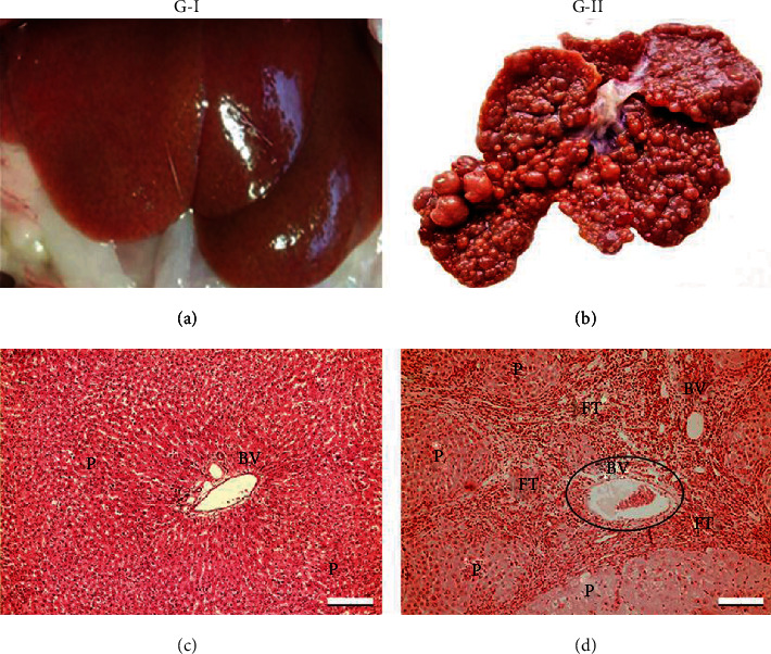
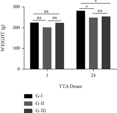
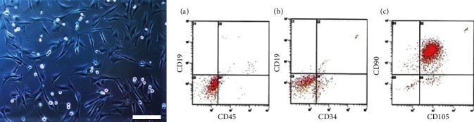
Hepatic fibrosis is caused by chronic injury due to toxic, infectious, or metabolic causes, and it may progress to cirrhosis and hepatocellular carcinoma. There is currently no antifibrotic therapy authorized for human use; however, there are promising studies using cell therapies. There are also no animal models that exactly reproduce human liver fibrosis that can be used to better understand the mechanisms of its regression and identify new targets for treatment and therapeutic approaches. On the other hand, mesenchymal stem cells (MSC) have experimentally demonstrated fibrosis regression effects, but it is necessary to have an animal model of advanced liver fibrosis to evaluate the effect of these cells. The aim of this work was to establish a protocol for the induction of advanced liver fibrosis in rats using thioacetamide (TAA), which will allow us to perform trials using MSC as a possible therapy for fibrosis regression. For this purpose, we selected 24 female rats and grouped them into three experimental groups: the control group (G-I) without treatment and groups II (G-II) and III (G-III) that received TAA by intraperitoneal injection for 24 weeks. Then, 1 × 10⁶/kg adipose mesenchymal stem cells (ASCs) were infused intravenously. Groups G-I and G-II were sacrificed 7 days after the last dose of ASC, and G-III was sacrificed 8 weeks after the last ASC infusion, all with xylazine/ketamine (40 mg/kg). The protocol used in this work established a model of advanced hepatic fibrosis as corroborated by METAVIR tests of the histological lesions; by the high levels of the markers α-SMA, CD68, and collagen type I; by functional alterations due to elevated markers of the hepatic lesions; and by alterations of the leukocytes, lymphocytes, and platelets. Finally, transplanted cells in the fibrous liver were detected. We conclude that TAA applied using the protocol introduced in this study induces a good model of advanced liver fibrosis in rats.


 分享
分享

 求助内容:
求助内容: 应助结果提醒方式:
 扫码关注我们
