Shin-Ichiro Hagiwara, Shigeharu Ueki, Ken Watanabe, Keinosuke Hizuka, Yuri Etani
{"title":"高嗜酸性粒细胞综合征累及胃肠道1例,表现为组织嗜酸性粒细胞溶解。","authors":"Shin-Ichiro Hagiwara, Shigeharu Ueki, Ken Watanabe, Keinosuke Hizuka, Yuri Etani","doi":"10.5415/apallergy.2022.12.e37","DOIUrl":null,"url":null,"abstract":"<p><p>Hypereosinophilic syndrome (HES), which is characterized by eosinophilia in the peripheral blood, often causes various organ disorders, including those of the gastrointestinal (GI) tract. The eosinophils play a key role in inflammation in eosinophilic GI disorders (EGIDs), including HES with GI involvement. Here, we report a case of HES with GI involvement that showed major basic proteins (MBPs) deposition in the absence of marked eosinophilic infiltration in the mucosa of the GI tract. An 11-year-old boy presented with nausea and epigastric pain for one week. He had a history of idiopathic HES with eosinophilic cystitis, diagnosed at the age of 2 years. He had been taking a low dose of corticosteroids for 9 years. The peripheral blood eosinophil count was 2,254/μL. Endoscopy revealed a swelling of the duodenal bulb mucosa. Histological findings of the duodenal mucosa revealed chronic inflammation, but no evidence of significant eosinophil infiltration and we could not diagnose him with HES with GI involvement or EGID. Immunofluorescent staining for MBP and galectin-10 was performed to detect intact and cytolytic eosinophils (eosinophil extracellular trap cell death: EETosis). Marked MBP deposition was evident in a small number of intact eosinophils in tissues from the duodenum, gastric antrum, and terminal ileum. The current case illustrates the utility of immunostaining for the detection of persistent eosinophilic inflammation, especially when cytolytic eosinophils are dominant.</p>","PeriodicalId":8488,"journal":{"name":"Asia Pacific Allergy","volume":"12 4","pages":"e37"},"PeriodicalIF":2.1000,"publicationDate":"2022-10-17","publicationTypes":"Journal Article","fieldsOfStudy":null,"isOpenAccess":false,"openAccessPdf":"https://ftp.ncbi.nlm.nih.gov/pub/pmc/oa_pdf/8f/ed/apa-12-e37.PMC9669462.pdf","citationCount":"2","resultStr":"{\"title\":\"Case of hypereosinophilic syndrome with gastrointestinal involvement showing tissue eosinophil cytolysis.\",\"authors\":\"Shin-Ichiro Hagiwara, Shigeharu Ueki, Ken Watanabe, Keinosuke Hizuka, Yuri Etani\",\"doi\":\"10.5415/apallergy.2022.12.e37\",\"DOIUrl\":null,\"url\":null,\"abstract\":\"<p><p>Hypereosinophilic syndrome (HES), which is characterized by eosinophilia in the peripheral blood, often causes various organ disorders, including those of the gastrointestinal (GI) tract. The eosinophils play a key role in inflammation in eosinophilic GI disorders (EGIDs), including HES with GI involvement. Here, we report a case of HES with GI involvement that showed major basic proteins (MBPs) deposition in the absence of marked eosinophilic infiltration in the mucosa of the GI tract. An 11-year-old boy presented with nausea and epigastric pain for one week. He had a history of idiopathic HES with eosinophilic cystitis, diagnosed at the age of 2 years. He had been taking a low dose of corticosteroids for 9 years. The peripheral blood eosinophil count was 2,254/μL. Endoscopy revealed a swelling of the duodenal bulb mucosa. Histological findings of the duodenal mucosa revealed chronic inflammation, but no evidence of significant eosinophil infiltration and we could not diagnose him with HES with GI involvement or EGID. Immunofluorescent staining for MBP and galectin-10 was performed to detect intact and cytolytic eosinophils (eosinophil extracellular trap cell death: EETosis). Marked MBP deposition was evident in a small number of intact eosinophils in tissues from the duodenum, gastric antrum, and terminal ileum. The current case illustrates the utility of immunostaining for the detection of persistent eosinophilic inflammation, especially when cytolytic eosinophils are dominant.</p>\",\"PeriodicalId\":8488,\"journal\":{\"name\":\"Asia Pacific Allergy\",\"volume\":\"12 4\",\"pages\":\"e37\"},\"PeriodicalIF\":2.1000,\"publicationDate\":\"2022-10-17\",\"publicationTypes\":\"Journal Article\",\"fieldsOfStudy\":null,\"isOpenAccess\":false,\"openAccessPdf\":\"https://ftp.ncbi.nlm.nih.gov/pub/pmc/oa_pdf/8f/ed/apa-12-e37.PMC9669462.pdf\",\"citationCount\":\"2\",\"resultStr\":null,\"platform\":\"Semanticscholar\",\"paperid\":null,\"PeriodicalName\":\"Asia Pacific Allergy\",\"FirstCategoryId\":\"1085\",\"ListUrlMain\":\"https://doi.org/10.5415/apallergy.2022.12.e37\",\"RegionNum\":0,\"RegionCategory\":null,\"ArticlePicture\":[],\"TitleCN\":null,\"AbstractTextCN\":null,\"PMCID\":null,\"EPubDate\":\"2022/10/1 0:00:00\",\"PubModel\":\"eCollection\",\"JCR\":\"Q3\",\"JCRName\":\"ALLERGY\",\"Score\":null,\"Total\":0}","platform":"Semanticscholar","paperid":null,"PeriodicalName":"Asia Pacific Allergy","FirstCategoryId":"1085","ListUrlMain":"https://doi.org/10.5415/apallergy.2022.12.e37","RegionNum":0,"RegionCategory":null,"ArticlePicture":[],"TitleCN":null,"AbstractTextCN":null,"PMCID":null,"EPubDate":"2022/10/1 0:00:00","PubModel":"eCollection","JCR":"Q3","JCRName":"ALLERGY","Score":null,"Total":0}
Case of hypereosinophilic syndrome with gastrointestinal involvement showing tissue eosinophil cytolysis.
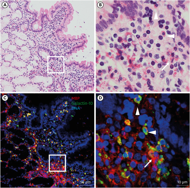
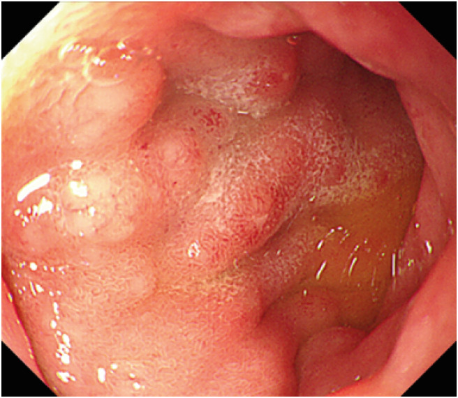
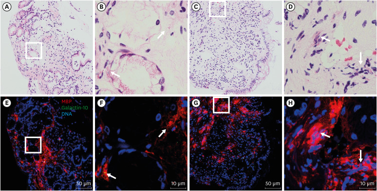
Hypereosinophilic syndrome (HES), which is characterized by eosinophilia in the peripheral blood, often causes various organ disorders, including those of the gastrointestinal (GI) tract. The eosinophils play a key role in inflammation in eosinophilic GI disorders (EGIDs), including HES with GI involvement. Here, we report a case of HES with GI involvement that showed major basic proteins (MBPs) deposition in the absence of marked eosinophilic infiltration in the mucosa of the GI tract. An 11-year-old boy presented with nausea and epigastric pain for one week. He had a history of idiopathic HES with eosinophilic cystitis, diagnosed at the age of 2 years. He had been taking a low dose of corticosteroids for 9 years. The peripheral blood eosinophil count was 2,254/μL. Endoscopy revealed a swelling of the duodenal bulb mucosa. Histological findings of the duodenal mucosa revealed chronic inflammation, but no evidence of significant eosinophil infiltration and we could not diagnose him with HES with GI involvement or EGID. Immunofluorescent staining for MBP and galectin-10 was performed to detect intact and cytolytic eosinophils (eosinophil extracellular trap cell death: EETosis). Marked MBP deposition was evident in a small number of intact eosinophils in tissues from the duodenum, gastric antrum, and terminal ileum. The current case illustrates the utility of immunostaining for the detection of persistent eosinophilic inflammation, especially when cytolytic eosinophils are dominant.
期刊介绍:
Asia Pacific Allergy (AP Allergy) is the official journal of the Asia Pacific Association of Allergy, Asthma and Clinical Immunology (APAAACI). Although the primary aim of the journal is to promote communication between Asia Pacific scientists who are interested in allergy, asthma, and clinical immunology including immunodeficiency, the journal is intended to be available worldwide. To enable scientists and clinicians from emerging societies appreciate the scope and intent of the journal, early issues will contain more educational review material. For better communication and understanding, it will include rational concepts related to the diagnosis and management of asthma and other immunological conditions. Over time, the journal will increase the number of original research papers to become the foremost citation journal for allergy and clinical immunology information of the Asia Pacific in the future.


 分享
分享
 求助内容:
求助内容: 应助结果提醒方式:
 扫码关注我们
