{"title":"56岁糖尿病患者,伴有急性胃脘痛。","authors":"Mehran Sotoodehnia, Arash Safaie","doi":"10.22114/AJEM.v0i0.49","DOIUrl":null,"url":null,"abstract":"LEARNING POINTS: Pathologic findings There is air in the wall/lumen of the gallbladder seen as multiple round or linear lucencies (yellow arrows in figure 2A and 2C). Mural enhancement of gallbladder is not seen in this oral and intravenous contrast-enhanced abdominal computed tomography (CT) scan. The gallbladder wall is irregular, and intraluminal membranes can be seen as irregular intraluminal linear and soft-tissue densities (black arrows in figure 2C). No contrast material can be seen inside the gallbladder. An abnormal loculated and encapsulated fluid attenuation adjacent to the gallbladder consistent with a pericholecystic abscess is another finding in this imaging [shown in figure 2D as (a)]. The gallstones which are seen as hyperdensities within the gallbladder lumen (shown with blue arrows in figure 2B and 2D), pericholecystic fat stranding","PeriodicalId":7290,"journal":{"name":"Advanced Journal of Emergency Medicine","volume":"2 2","pages":"e24"},"PeriodicalIF":0.0000,"publicationDate":"2018-01-21","publicationTypes":"Journal Article","fieldsOfStudy":null,"isOpenAccess":false,"openAccessPdf":"https://ftp.ncbi.nlm.nih.gov/pub/pmc/oa_pdf/f5/ca/AJEM-2-e24.PMC6549057.pdf","citationCount":"0","resultStr":"{\"title\":\"A 56-year-old Diabetic Man with Acute Epigastric Pain.\",\"authors\":\"Mehran Sotoodehnia, Arash Safaie\",\"doi\":\"10.22114/AJEM.v0i0.49\",\"DOIUrl\":null,\"url\":null,\"abstract\":\"LEARNING POINTS: Pathologic findings There is air in the wall/lumen of the gallbladder seen as multiple round or linear lucencies (yellow arrows in figure 2A and 2C). Mural enhancement of gallbladder is not seen in this oral and intravenous contrast-enhanced abdominal computed tomography (CT) scan. The gallbladder wall is irregular, and intraluminal membranes can be seen as irregular intraluminal linear and soft-tissue densities (black arrows in figure 2C). No contrast material can be seen inside the gallbladder. An abnormal loculated and encapsulated fluid attenuation adjacent to the gallbladder consistent with a pericholecystic abscess is another finding in this imaging [shown in figure 2D as (a)]. The gallstones which are seen as hyperdensities within the gallbladder lumen (shown with blue arrows in figure 2B and 2D), pericholecystic fat stranding\",\"PeriodicalId\":7290,\"journal\":{\"name\":\"Advanced Journal of Emergency Medicine\",\"volume\":\"2 2\",\"pages\":\"e24\"},\"PeriodicalIF\":0.0000,\"publicationDate\":\"2018-01-21\",\"publicationTypes\":\"Journal Article\",\"fieldsOfStudy\":null,\"isOpenAccess\":false,\"openAccessPdf\":\"https://ftp.ncbi.nlm.nih.gov/pub/pmc/oa_pdf/f5/ca/AJEM-2-e24.PMC6549057.pdf\",\"citationCount\":\"0\",\"resultStr\":null,\"platform\":\"Semanticscholar\",\"paperid\":null,\"PeriodicalName\":\"Advanced Journal of Emergency Medicine\",\"FirstCategoryId\":\"1085\",\"ListUrlMain\":\"https://doi.org/10.22114/AJEM.v0i0.49\",\"RegionNum\":0,\"RegionCategory\":null,\"ArticlePicture\":[],\"TitleCN\":null,\"AbstractTextCN\":null,\"PMCID\":null,\"EPubDate\":\"2018/1/1 0:00:00\",\"PubModel\":\"eCollection\",\"JCR\":\"\",\"JCRName\":\"\",\"Score\":null,\"Total\":0}","platform":"Semanticscholar","paperid":null,"PeriodicalName":"Advanced Journal of Emergency Medicine","FirstCategoryId":"1085","ListUrlMain":"https://doi.org/10.22114/AJEM.v0i0.49","RegionNum":0,"RegionCategory":null,"ArticlePicture":[],"TitleCN":null,"AbstractTextCN":null,"PMCID":null,"EPubDate":"2018/1/1 0:00:00","PubModel":"eCollection","JCR":"","JCRName":"","Score":null,"Total":0}
A 56-year-old Diabetic Man with Acute Epigastric Pain.
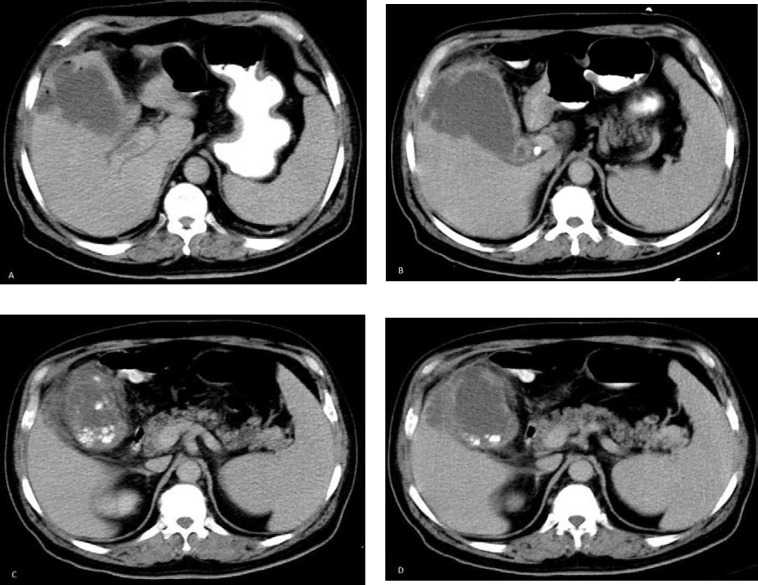
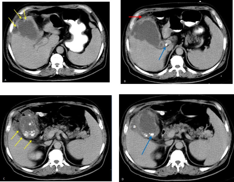
LEARNING POINTS: Pathologic findings There is air in the wall/lumen of the gallbladder seen as multiple round or linear lucencies (yellow arrows in figure 2A and 2C). Mural enhancement of gallbladder is not seen in this oral and intravenous contrast-enhanced abdominal computed tomography (CT) scan. The gallbladder wall is irregular, and intraluminal membranes can be seen as irregular intraluminal linear and soft-tissue densities (black arrows in figure 2C). No contrast material can be seen inside the gallbladder. An abnormal loculated and encapsulated fluid attenuation adjacent to the gallbladder consistent with a pericholecystic abscess is another finding in this imaging [shown in figure 2D as (a)]. The gallstones which are seen as hyperdensities within the gallbladder lumen (shown with blue arrows in figure 2B and 2D), pericholecystic fat stranding


 分享
分享

 求助内容:
求助内容: 应助结果提醒方式:
 扫码关注我们
