{"title":"恶性间皮瘤的细胞学诊断:一个病例系列。","authors":"Sakshi Dahiya, Meeta Singh, Shyama Jain, Bembem Khuraijam, Naman Suroya, Shramana Mandal","doi":"10.4103/joc.joc_145_21","DOIUrl":null,"url":null,"abstract":"<p><strong>Background: </strong>Mesotheliomas are neoplasms of the serosal lining of the body cavities. Diagnosis requires a multimodal approach of clinical findings, cytology, and histopathology with immunohistochemistry (IHC). The published sensitivity of cytology for diagnosing mesothelioma ranges from 30% to 75%.</p><p><strong>Aim and objectives: </strong>This study aimed to calculate the incidence of malignant mesothelioma (MM) at our institute and to study the cytological features of MM.</p><p><strong>Materials and methods: </strong>A retrospective study of pleural, peritoneal, and pericardial fluids submitted at our institute was done. The duration of the study was 8 years (2011-2019). Apart from examining Giemsa smears, a panel of immunocytochemical (ICC) and cell block immunohistochemical (IHC) markers was applied to achieve the diagnosis. These included calretinin, mesothelin, CK5/6, Hector Battifora mesothelial cell antibody (HBME), WT1, MOC31, CK7 and CK20. Histopathological correlation was done wherever possible.</p><p><strong>Result: </strong>In the present study, we compiled four cases of MM over 8 years diagnosed on serous effusion cytology and confirmed by immunocytochemistry (ICC)/cell block immunohistochemistry (IHC)/biopsy. This indicates a rare incidence of MM. The Cytological features of MM were studied.</p><p><strong>Conclusion: </strong>The diagnosis of MM is difficult, especially cytologically. It was found to be a rare entity in the malignant cases diagnosed on effusion cytology.</p>","PeriodicalId":50217,"journal":{"name":"Journal of Cytology","volume":null,"pages":null},"PeriodicalIF":1.0000,"publicationDate":"2022-07-01","publicationTypes":"Journal Article","fieldsOfStudy":null,"isOpenAccess":false,"openAccessPdf":"https://www.ncbi.nlm.nih.gov/pmc/articles/PMC9585811/pdf/","citationCount":"1","resultStr":"{\"title\":\"Cytological Diagnosis of Malignant Mesothelioma: A Case Series.\",\"authors\":\"Sakshi Dahiya, Meeta Singh, Shyama Jain, Bembem Khuraijam, Naman Suroya, Shramana Mandal\",\"doi\":\"10.4103/joc.joc_145_21\",\"DOIUrl\":null,\"url\":null,\"abstract\":\"<p><strong>Background: </strong>Mesotheliomas are neoplasms of the serosal lining of the body cavities. Diagnosis requires a multimodal approach of clinical findings, cytology, and histopathology with immunohistochemistry (IHC). The published sensitivity of cytology for diagnosing mesothelioma ranges from 30% to 75%.</p><p><strong>Aim and objectives: </strong>This study aimed to calculate the incidence of malignant mesothelioma (MM) at our institute and to study the cytological features of MM.</p><p><strong>Materials and methods: </strong>A retrospective study of pleural, peritoneal, and pericardial fluids submitted at our institute was done. The duration of the study was 8 years (2011-2019). Apart from examining Giemsa smears, a panel of immunocytochemical (ICC) and cell block immunohistochemical (IHC) markers was applied to achieve the diagnosis. These included calretinin, mesothelin, CK5/6, Hector Battifora mesothelial cell antibody (HBME), WT1, MOC31, CK7 and CK20. Histopathological correlation was done wherever possible.</p><p><strong>Result: </strong>In the present study, we compiled four cases of MM over 8 years diagnosed on serous effusion cytology and confirmed by immunocytochemistry (ICC)/cell block immunohistochemistry (IHC)/biopsy. This indicates a rare incidence of MM. The Cytological features of MM were studied.</p><p><strong>Conclusion: </strong>The diagnosis of MM is difficult, especially cytologically. It was found to be a rare entity in the malignant cases diagnosed on effusion cytology.</p>\",\"PeriodicalId\":50217,\"journal\":{\"name\":\"Journal of Cytology\",\"volume\":null,\"pages\":null},\"PeriodicalIF\":1.0000,\"publicationDate\":\"2022-07-01\",\"publicationTypes\":\"Journal Article\",\"fieldsOfStudy\":null,\"isOpenAccess\":false,\"openAccessPdf\":\"https://www.ncbi.nlm.nih.gov/pmc/articles/PMC9585811/pdf/\",\"citationCount\":\"1\",\"resultStr\":null,\"platform\":\"Semanticscholar\",\"paperid\":null,\"PeriodicalName\":\"Journal of Cytology\",\"FirstCategoryId\":\"3\",\"ListUrlMain\":\"https://doi.org/10.4103/joc.joc_145_21\",\"RegionNum\":4,\"RegionCategory\":\"医学\",\"ArticlePicture\":[],\"TitleCN\":null,\"AbstractTextCN\":null,\"PMCID\":null,\"EPubDate\":\"2022/7/30 0:00:00\",\"PubModel\":\"Epub\",\"JCR\":\"Q4\",\"JCRName\":\"MEDICAL LABORATORY TECHNOLOGY\",\"Score\":null,\"Total\":0}","platform":"Semanticscholar","paperid":null,"PeriodicalName":"Journal of Cytology","FirstCategoryId":"3","ListUrlMain":"https://doi.org/10.4103/joc.joc_145_21","RegionNum":4,"RegionCategory":"医学","ArticlePicture":[],"TitleCN":null,"AbstractTextCN":null,"PMCID":null,"EPubDate":"2022/7/30 0:00:00","PubModel":"Epub","JCR":"Q4","JCRName":"MEDICAL LABORATORY TECHNOLOGY","Score":null,"Total":0}
Cytological Diagnosis of Malignant Mesothelioma: A Case Series.
Background: Mesotheliomas are neoplasms of the serosal lining of the body cavities. Diagnosis requires a multimodal approach of clinical findings, cytology, and histopathology with immunohistochemistry (IHC). The published sensitivity of cytology for diagnosing mesothelioma ranges from 30% to 75%.
Aim and objectives: This study aimed to calculate the incidence of malignant mesothelioma (MM) at our institute and to study the cytological features of MM.
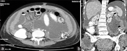
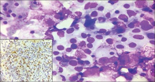
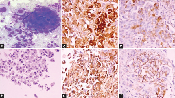
Materials and methods: A retrospective study of pleural, peritoneal, and pericardial fluids submitted at our institute was done. The duration of the study was 8 years (2011-2019). Apart from examining Giemsa smears, a panel of immunocytochemical (ICC) and cell block immunohistochemical (IHC) markers was applied to achieve the diagnosis. These included calretinin, mesothelin, CK5/6, Hector Battifora mesothelial cell antibody (HBME), WT1, MOC31, CK7 and CK20. Histopathological correlation was done wherever possible.
Result: In the present study, we compiled four cases of MM over 8 years diagnosed on serous effusion cytology and confirmed by immunocytochemistry (ICC)/cell block immunohistochemistry (IHC)/biopsy. This indicates a rare incidence of MM. The Cytological features of MM were studied.
Conclusion: The diagnosis of MM is difficult, especially cytologically. It was found to be a rare entity in the malignant cases diagnosed on effusion cytology.
期刊介绍:
The Journal of Cytology is the official Quarterly publication of the Indian Academy of Cytologists. It is in the 25th year of publication in the year 2008. The journal covers all aspects of diagnostic cytology, including fine needle aspiration cytology, gynecological and non-gynecological cytology. Articles on ancillary techniques, like cytochemistry, immunocytochemistry, electron microscopy, molecular cytopathology, as applied to cytological material are also welcome. The journal gives preference to clinically oriented studies over experimental and animal studies. The Journal would publish peer-reviewed original research papers, case reports, systematic reviews, meta-analysis, and debates.


 分享
分享
 求助内容:
求助内容: 应助结果提醒方式:
 扫码关注我们
