Bahare Saidi, Babak Fallahi, Armaghan Fard-Esfahani, Alireza Emami-Ardekani, Mohammad Eftekhari
{"title":"非皮肤黑色素瘤,FDG PET/CT的表现和预后价值:23例患者的病例系列和文献综述。","authors":"Bahare Saidi, Babak Fallahi, Armaghan Fard-Esfahani, Alireza Emami-Ardekani, Mohammad Eftekhari","doi":"10.22038/AOJNMB.2022.61517.1433","DOIUrl":null,"url":null,"abstract":"<p><strong>Objectives: </strong>Non-cutaneous malignant melanomas (NCM) are rare malignancies. Due to their nonspecific symptoms, they present later in life. The value of FDG PET/CT in this group of patients is not clear. The aim of this study is to assess the role of FDG PET/CT in the management of NCM and its prognostic implication.</p><p><strong>Methods: </strong>We retrospectively selected twenty-three patients with a diagnosis of NCM evaluated with FDG PET/CT in Shariati hospital between 2019 and 2021. The PET/CT data were reviewed and compared with available conventional imaging findings. Five patients died within five months. The surviving patients were followed within a time interval of 7 to 27.5 months after their PET/CT study, regarding their disease status.</p><p><strong>Results: </strong>Among 23 patients (8 ocular, 5 sinonasal, 3 pharyngeal, 2 anorectal, 2 vulvovaginal, and 3 unknown primaries), PET/CT was able to detect residual primary disease, assess treatment response, and reveal or exclude metastases. Additional lesions compared to conventional imaging were found in five, while in one with brain metastases PET/CT was unable to detect lesions on MRI. Thirteen patients had negative PET/CT finding of which 11 (85%) did not have remarkable finding on follow-up. Metastatic disease was recognized in eight. Patients with extensive metastases on FDG PET/CT had a poorer outcome.</p><p><strong>Conclusion: </strong>Similar to cutaneous melanoma, PET/CT is valuable in the management of NCM patients and is superior to conventional imaging modalities, with the exception of brain metastases. Patients with negative PET/CT findings have a better outcome as opposed to patients with significant positive PET/CT findings.</p>","PeriodicalId":8503,"journal":{"name":"Asia Oceania Journal of Nuclear Medicine and Biology","volume":null,"pages":null},"PeriodicalIF":0.0000,"publicationDate":"2022-01-01","publicationTypes":"Journal Article","fieldsOfStudy":null,"isOpenAccess":false,"openAccessPdf":"https://www.ncbi.nlm.nih.gov/pmc/articles/PMC9205851/pdf/","citationCount":"1","resultStr":"{\"title\":\"Non-Cutaneous Melanoma, Findings and Prognostic Value of FDG PET/CT: A Case Series of 23 patients and review of the literature.\",\"authors\":\"Bahare Saidi, Babak Fallahi, Armaghan Fard-Esfahani, Alireza Emami-Ardekani, Mohammad Eftekhari\",\"doi\":\"10.22038/AOJNMB.2022.61517.1433\",\"DOIUrl\":null,\"url\":null,\"abstract\":\"<p><strong>Objectives: </strong>Non-cutaneous malignant melanomas (NCM) are rare malignancies. Due to their nonspecific symptoms, they present later in life. The value of FDG PET/CT in this group of patients is not clear. The aim of this study is to assess the role of FDG PET/CT in the management of NCM and its prognostic implication.</p><p><strong>Methods: </strong>We retrospectively selected twenty-three patients with a diagnosis of NCM evaluated with FDG PET/CT in Shariati hospital between 2019 and 2021. The PET/CT data were reviewed and compared with available conventional imaging findings. Five patients died within five months. The surviving patients were followed within a time interval of 7 to 27.5 months after their PET/CT study, regarding their disease status.</p><p><strong>Results: </strong>Among 23 patients (8 ocular, 5 sinonasal, 3 pharyngeal, 2 anorectal, 2 vulvovaginal, and 3 unknown primaries), PET/CT was able to detect residual primary disease, assess treatment response, and reveal or exclude metastases. Additional lesions compared to conventional imaging were found in five, while in one with brain metastases PET/CT was unable to detect lesions on MRI. Thirteen patients had negative PET/CT finding of which 11 (85%) did not have remarkable finding on follow-up. Metastatic disease was recognized in eight. Patients with extensive metastases on FDG PET/CT had a poorer outcome.</p><p><strong>Conclusion: </strong>Similar to cutaneous melanoma, PET/CT is valuable in the management of NCM patients and is superior to conventional imaging modalities, with the exception of brain metastases. Patients with negative PET/CT findings have a better outcome as opposed to patients with significant positive PET/CT findings.</p>\",\"PeriodicalId\":8503,\"journal\":{\"name\":\"Asia Oceania Journal of Nuclear Medicine and Biology\",\"volume\":null,\"pages\":null},\"PeriodicalIF\":0.0000,\"publicationDate\":\"2022-01-01\",\"publicationTypes\":\"Journal Article\",\"fieldsOfStudy\":null,\"isOpenAccess\":false,\"openAccessPdf\":\"https://www.ncbi.nlm.nih.gov/pmc/articles/PMC9205851/pdf/\",\"citationCount\":\"1\",\"resultStr\":null,\"platform\":\"Semanticscholar\",\"paperid\":null,\"PeriodicalName\":\"Asia Oceania Journal of Nuclear Medicine and Biology\",\"FirstCategoryId\":\"1085\",\"ListUrlMain\":\"https://doi.org/10.22038/AOJNMB.2022.61517.1433\",\"RegionNum\":0,\"RegionCategory\":null,\"ArticlePicture\":[],\"TitleCN\":null,\"AbstractTextCN\":null,\"PMCID\":null,\"EPubDate\":\"\",\"PubModel\":\"\",\"JCR\":\"Q3\",\"JCRName\":\"Medicine\",\"Score\":null,\"Total\":0}","platform":"Semanticscholar","paperid":null,"PeriodicalName":"Asia Oceania Journal of Nuclear Medicine and Biology","FirstCategoryId":"1085","ListUrlMain":"https://doi.org/10.22038/AOJNMB.2022.61517.1433","RegionNum":0,"RegionCategory":null,"ArticlePicture":[],"TitleCN":null,"AbstractTextCN":null,"PMCID":null,"EPubDate":"","PubModel":"","JCR":"Q3","JCRName":"Medicine","Score":null,"Total":0}
Non-Cutaneous Melanoma, Findings and Prognostic Value of FDG PET/CT: A Case Series of 23 patients and review of the literature.
Objectives: Non-cutaneous malignant melanomas (NCM) are rare malignancies. Due to their nonspecific symptoms, they present later in life. The value of FDG PET/CT in this group of patients is not clear. The aim of this study is to assess the role of FDG PET/CT in the management of NCM and its prognostic implication.
Methods: We retrospectively selected twenty-three patients with a diagnosis of NCM evaluated with FDG PET/CT in Shariati hospital between 2019 and 2021. The PET/CT data were reviewed and compared with available conventional imaging findings. Five patients died within five months. The surviving patients were followed within a time interval of 7 to 27.5 months after their PET/CT study, regarding their disease status.
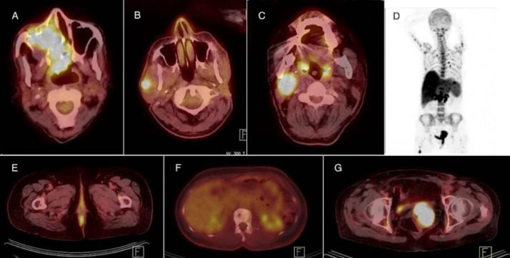
Results: Among 23 patients (8 ocular, 5 sinonasal, 3 pharyngeal, 2 anorectal, 2 vulvovaginal, and 3 unknown primaries), PET/CT was able to detect residual primary disease, assess treatment response, and reveal or exclude metastases. Additional lesions compared to conventional imaging were found in five, while in one with brain metastases PET/CT was unable to detect lesions on MRI. Thirteen patients had negative PET/CT finding of which 11 (85%) did not have remarkable finding on follow-up. Metastatic disease was recognized in eight. Patients with extensive metastases on FDG PET/CT had a poorer outcome.
Conclusion: Similar to cutaneous melanoma, PET/CT is valuable in the management of NCM patients and is superior to conventional imaging modalities, with the exception of brain metastases. Patients with negative PET/CT findings have a better outcome as opposed to patients with significant positive PET/CT findings.


 分享
分享
 求助内容:
求助内容: 应助结果提醒方式:
 扫码关注我们
